Veterinary specialists have utilized organoids—three-layered organ-like designs developed from immature microorganisms and tissue tests—to examine the natural cycles of cellular breakdown in the lungs in canines, an illness that is a lot more extraordinary in our canine companions than it is in people, yet at the same time frequently undeniably more lethal.
Organoids are three-layered organ-like designs filled in the lab that propose far more prominent constancy to complex natural cycles than customary cell societies developed on level 2D surfaces. These state-of-the-art models have, as of late, been formed to help investigate human science and sickness. Yet, specialists have now interestingly applied the examination method to cellular breakdown in the lungs in canines.
A review depicting the scientists’ methods and discoveries was distributed in the journal Biomedicine and Pharmacotherapy.
“The expectation was that these organoids would more accurately mimic the tissue architecture of canine lung cancer than any prior cell culture attempts.”
Tatsuya Usui corresponding author of the study and Associate Professor at the Division of Animal Life Science.
In the domain of malignant growth research, cellular breakdown in the lungs has consistently lingered as a considerable enemy in the human world; however, our cherished canine colleagues can likewise experience the ill effects of their own rendition of this sickness, known as canine essential cellular breakdown in the lungs.
For canines determined to have cellular breakdown in the lungs, the viewpoint can be dreary. Many cases are possibly found when the infection has advanced, essentially prompting restricted treatment choices. Careful evacuation of lung tissue, known as lung lobectomy, is a typical methodology, yet it may not be healing for cutting-edge cases.
Truth be told, the middle endurance time for canines going through lung lobectomy floats somewhere in the range of 160 and 450 days, even with chemotherapy. For canines with broad disease development, the visualization is considerably more dreary, with endurance times going from a simple 60 to 180 days.
In contrast to human medication, where sub-atomically designated treatments have turned into a unique advantage, such medicines are rare in veterinary work, leaving our fuzzy companions with restricted choices.
Enter the universe of 3D organoid societies, a state-of-the-art innovation that emulates the elements of living tissue better compared to customary cell societies. 3D organoids are smaller than expected, three-layered structures that are refined from undifferentiated cells or tissue tests in a lab. The undifferentiated cells or tissue tests can self-assemble into complex, organ-like designs, thus “organoid.”
The organoid’s cells and designs can collaborate with one another very much like a genuine organ in a living organic entity in three aspects, rather than conventional cell societies developed on level, 2D surfaces. This permits researchers to concentrate on substantially more intricate natural cycles.
The veterinary medication specialists believed that they could apply this new strategy to the prickly issue of canine cellular breakdown in the lungs with the expectation that they could recognize novel remedial routes.
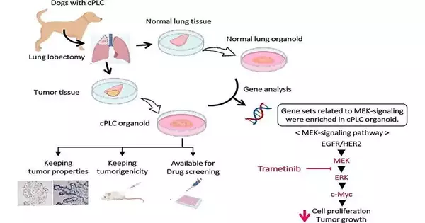
They took tests of growths from canines with cellular breakdown in the lungs along with the solid pieces of their lungs to make canine essential cellular breakdown in the lungs organoids (cPLCO) and canine typical lung organoids (cNLO).
“The expectation was that these organoids would reproduce the tissue design of canine cellular breakdown in the lungs better than any past endeavors at cell societies,” said Tatsuya Usui, comparing the creator of the review and academic administrator at the Division of Creature Life Science with the Tokyo College of Horticulture and Innovation.
The analysts found that the cPLCO loyally reflected the attributes of their unique growth tissues, both in histological morphology (fundamentally, the shape and design of the tissues) and atomic profiles, including the outflow of explicit cancer markers. Growth markers are any particles, normally proteins, created by the disease cells or by different cells in the body because of the presence of a malignant growth that give any data about the malignant growth.
These markers, created in higher amounts than in ordinary solid patients, are what clinicians and analysts use to recognize how forceful the disease is and whether it is responding to treatment.
They found that various kinds of canine cellular breakdowns in the lungs displayed differing aversions to disease drugs, paving the way for the potential for customized treatment.
The researchers likewise distinguished a relationship between the reasonability of organoid cells and the articulation levels of explicit objective particles (fundamentally, the way that frequently a quality gets “turned on, for example, Human Epidermal Development Element Receptor 2 (HER2) and Epidermal Development Variable Receptor (EGFR). These are the two proteins that assume urgent roles in cell flagging, development, and guidelines. This perception could make it ready for treatments that focus on these atoms later on.
Sequencing of RNA uncovered that the harmful organoid showed huge expansion in the “turning on” of 11 qualities related to growth multiplication in different diseases.
Maybe most tantalizingly, the malignant organoid showed enhancement in the Mitogen-Enacted Protein Kinase flagging pathway, likewise referred to all the more essentially as the MEK-flagging pathway, a promising road for mediation, as its sub-atomic cycles empower transmission of data from receptors on the outer layer of a cell to the DNA in its core.
This pathway, including an outpouring of proteins following up on different proteins, thus following up on different proteins, etc., is vital to cell multiplication, separation, endurance, and answering data signals from outside the phone. Frequently, assuming that any of the means on the cycle stall out in an on or off position, it can bring about cancer development.
The scientists tried a MEK inhibitor called trametinib, which essentially diminished the suitability of the malignant organoid and repressed growth development when disease cells were united into solid cell models.
The production of canine cellular breakdown in the lungs organoids is an incredible asset to investigating the sickness in uncommon detail, possibly prompting more compelling therapies for our four-legged companions. However, we may try and expand its impact into the domain of human cellular breakdown in the lungs, offering new demonstrative markers and helpful targets.
More information: Yomogi Shiota (Sato) et al, Derivation of a new model of lung adenocarcinoma using canine lung cancer organoids for translational research in pulmonary medicine, Biomedicine & Pharmacotherapy (2023). DOI: 10.1016/j.biopha.2023.115079


