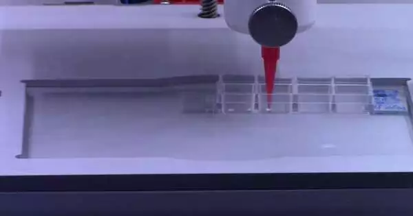
Another review from North Carolina State College shows a reproducible approach to concentrating on cell correspondence among shifted sorts of plant cells by “bioprinting” these cells through a 3D printer. Studying how plant cells communicate with one another — and with their surroundings — is critical to understanding how plant cells work and may eventually lead to better yield assortments and ideal developing conditions.
The analysts bioprinted cells from the model plant Arabidopsis thaliana and from soybeans to concentrate on not simply whether plant cells would live subsequent to being bioprinted—and for how long—but in addition to looking at how they acquire and change their character and capability.
“A plant root has many cell types with specific capabilities,” said Lisa Van Lair Broeck, a NC State postdoctoral scientist who is the main creator of a paper depicting the work. “Likewise, various arrangements of quality are being communicated; some are cell-explicit. We needed to realize what occurs after you bioprint live cells and spot them into a climate that you configuration: Would they say they are alive and doing what they ought to do? “
The course of 3D bioprinting plant cells is precisely like printing ink or plastic, with a couple of vital changes.
“Instead of 3D printing ink or plastic, we use ‘bioink,’ or living plant cells. The mechanics of both processes are the same, with a few notable differences for plant cells: an ultraviolet filter to keep the environment sterile and multiple print heads (rather than just one) to print different bioinks at the same time.”
Van den Broeck
“Rather than 3D printing ink or plastic, we use ‘bioink,’ or living plant cells,” Van Cave Broeck said. “The repairmen are similar in the two cycles with a couple of eminent contrasts for plant cells: a bright channel used to keep the climate sterile and various print heads—as opposed to only one—to print different bioinks all the while.”
Live plant cells without cell walls, or protoplasts, were bioprinted alongside supplements, development chemicals, and a thickening specialist called agarose—a kelp-based compound. Agarose gives cells strength and a platform, like mortar that upholds blocks in the mass of a structure.
“We observed that it is basic to utilize a legitimate platform,” said Ross Sozzani, teacher of plant and microbial science at NC State and a co-creator of the paper. When you print the bioink, you want it to be fluid, yet when it emerges, it should be strong. Copying the common habitat helps keep cell signals and prompts happening as they would in soil. “
The exploration showed that the greater part of the 3D bioprinted cells were suitable and isolated over the long haul to shape microcalli, or little states of cells.
“We expected great suitability on the day the cells were bioprinted, yet we had never kept up with cells past a couple of hours subsequent to bioprinting, so we had no clue about what might happen days after the fact,” Van Lair Broeck said. “Comparable feasibility ranges are displayed after physically pipetting cells, so the 3D printing process doesn’t appear to do anything unsafe to cells.”
“This is a physically troublesome cycle, and 3D bioprinting controls the strain of the drops and the speed at which the drops are printed,” Sozzani said. “Bioprinting gives better access to high throughput handling and command over the design of the cells subsequent to bioprinting, for example, layers or honeycomb shapes.”
The analysts likewise bioprinted individual cells to test whether they could recover, or isolate and increase. The discoveries showed that Arabidopsis root and shoot cells required various mixes of supplements and platforms for ideal feasibility.
In the interim, over 40% of individual soybean early stage cells stayed viable for fourteen days subsequent to bioprinting and were furthermore isolated over the long run to frame microcalli.
“This demonstrates the way that 3D bioprinting can be helpful to concentrate on cell recovery in crop plants,” Sozzani said.
Finally, the analysts concentrated on the cell characteristics of the bioprinted cells. Arabidopsis root cells and undeveloped soybean cells are known for their high expansion rates and the absence of fixed characteristics. As such, similar to creatures or human immature microorganisms, these cells can become different cell types.
“We found that bioprinted cells can assume the character of immature microorganisms; they partition and develop and communicate explicit qualities,” Van Noord Broeck said. “At the point when you bioprint, you print an entire populace of cell types. We had the option to analyze the qualities communicated by individual cells after 3D bioprinting to see any progressions in cell character. “
The analysts intend to proceed with their work, concentrating on cell correspondence after 3D bioprinting, including at the single-cell level.
By and large, this study shows the strong capability of utilizing 3D bioprinting to recognize the ideal mixtures expected to help plant cell feasibility and correspondence in a controlled climate, Sozzani said.
The exploration shows up in Science Advances. Tim Horn, partner teacher of mechanical and aviation design at NC State, is a co-creator of the paper.
More information: Lisa Van den Broeck et al, Establishing a reproducible approach to study cellular functions of plants cells with 3D bioprinting, Science Advances (2022). DOI: 10.1126/sciadv.abp9906. www.science.org/doi/10.1126/sciadv.abp9906
Journal information: Science Advances


