Humans are naturally capable of separating information about others in their social circle based on their own requirements. For instance, if they require advice regarding a legal matter or emotional support regarding a personal matter, they might contact a friend who is a lawyer.
Although the neural mechanisms underlying this human ability to selectively separate social information about others based on the task at hand are well documented, they have not yet been discovered. A new paper by specialists at the College of Oxford, distributed in Neuron, divulges a brain process that could assume a vital role in this extraordinary mental and social capacity.
“In numerous situations, for instance, looking for a companion to assist with home fixes, we ought to zero in on individuals’ capacity to play out the job that needs to be done and overlook their capacity in different spaces,” Ali Mahmoodi, one of the specialists who did the review, told Clinical Xpress.
“In many situations, such as looking for a friend to help with home repairs, we should focus on people’s ability to perform the task at hand while ignoring their ability in other domains,”
Ali Mahmoodi, one of the researchers who carried out the study,
“In this instance, we are more concerned about our friend’s capacity to use a screwdriver or hammer than they are an accomplished neuroscientist.” “We researched how the cerebrum takes care of the issue of choosing and isolating the most significant aspect for correlation, utilizing a multi-layered social examination task.”
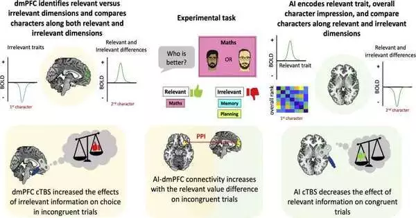
Mahmoodi and his colleagues conducted an experiment with 30 participants to investigate the neural mechanisms underlying the selection and separation of task-relevant social information. Using functional magnetic resonance imaging (fMRI), the brain activity of each of these participants was recorded as they completed a social comparison task. FMRI is a common imaging method used in neuroscience research to measure changes in blood flow linked to activity in various brain regions.
According to Mahmoodi, “We used an experimental paradigm in which people were required to compare two characters in one domain (relevant) while ignoring their ability in two other domains (irrelevant).” For instance, they needed to analyze their companions’ home fix capacity while disregarding how great they are at math and memory tests. In this paradigm, the key is to concentrate on the relevant trait (for instance, home repair skills) and ignore the two other irrelevant traits (for example, memory and math).
Mahmoodi and his colleagues discovered two brain regions that were particularly active as the participants completed the social comparison task when they examined the fMRI scans collected during their experiment. These two regions are the dorsomedial prefrontal cortex (dmPFC), which is known to play a crucial role in social cognition, and the anterior insula (AI), which controls the integration of subjective feelings into mental processes.
Mahmodi continued, “We found that people use irrelevant information to compare them when it is difficult for people to compare others in the relevant dimension (the two characters to be compared are similar).” We discovered a brain region in the medial prefrontal cortex that reduces the impact of irrelevant information on our decision-making at the neural level. Importantly, people’s comparisons of others were more influenced by irrelevant information when we temporarily disrupted this area’s activity using brain stimulation techniques than when we stimulated other brain areas.
This group of researchers recently discovered a neural circuit that may be responsible for the useful ability of humans to selectively sort through social information and focus on specific skills or qualities of others based on the context in which they are located. It might open the door to further research into the function of the dmPFC-AI circuit they discovered in the future.
More information: Ali Mahmoodi et al, Causal role of a neural system for separating and selecting multidimensional social cognitive information, Neuron (2023). DOI: 10.1016/j.neuron.2022.12.030


