Profound learning models have ended up being exceptionally encouraging devices for investigating huge quantities of pictures. Throughout the last ten years or something like that, they have, along these lines, been presented in an assortment of settings, including research labs.
In the area of science, profound learning models might actually work with the quantitative examination of microscopy pictures, permitting analysts to remove significant data from these pictures and decipher their perceptions. Preparing models to do this, in any case, can be a very challenging task, as it frequently requires the extraction of elements (i.e., number of cells, area of cells, and so forth) from microscopy pictures and the manual annotation of preparing information.
Specialists at CERVO Brain Research Center, the Institute for Intelligence and Data, and Université Laval in Canada have as of late fostered a counterfeit brain network that could act inside and out examinations of microscopy pictures utilizing more straightforward, picture-level explanations. This model, named MICRA-Net (MICRoscopy Analysis Brain Organization), was presented in a paper distributed in Nature Machine Intelligence.
“Our model’s lack of supervision is due to the way MICRA-Net is trained. The annotations needed to train MICRA-Net are basic binary (yes or no) classification labels that are significantly easier to get than sophisticated exact labels like contours of the structure of interest.”
Anthony Bilodeau, a Ph.D. student at Université Laval
“Physically extricating highlights from pictures is a long and monotonous errand, especially in cases where it should be performed by a prepared master,” Anthony Bilodeau, a Ph.D. understudy at Université Laval who did the review, told TechXplore. While profound learning (DL) models that include extraction are accessible, they actually require preparation with explanations, which are frequently difficult to acquire. Our model (MICRA-Net) depends on a straightforward characterization task, posing the inquiry: “is the construction present in the district of the picture that you are checking out or not?”
By resolving this basic inquiry, the model created by the group at Université Laval can foresee the presence or nonattendance of a particular design in pictures by utilizing straightforward double explanations. This significantly diminishes the time expected to clarify pictures and work on the preparation cycle, while as yet permitting the model to handle different microscopy picture investigation assignments all the while.
“Our model’s feeble management comes from how MICRA-Net is prepared,” Bilodeau said. “The explanations expected to prepare MICRA-Net are basic twofold (yes or no) characterization names, which are a lot simpler to get than complex exact marks, like shapes of the design of interest.”
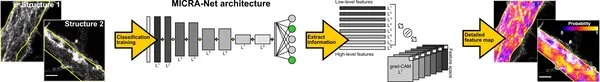
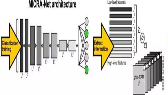
Conversely, with other existing profound learning instruments for the investigation of microscopy pictures, MICRA-Net can handle complex errands, like semantic division and identification, while utilizing far more straightforward, double picture explanations. It accomplishes this by extricating fundamental data about the design of interest from the slope class enacted maps (i.e., graduate CAMs).
“Consolidating the graduate CAMs of various layers of the organization permits the model to feature the construction of interest in the picture and can be utilized to produce exact division covers or to limit the articles,” Bilodeau made sense of. “MICRA-Net additionally accomplishes comparative or better execution on complex picture investigation errands contrasted with laid out baselines prepared utilizing powerless oversight (e.g., jumping box explanations, jots”).
In the underlying assessments conducted by the group at Université Laval, MICRA-Net accomplished wonderful outcomes, beating the vast majority of the models it was contrasted with. Later on, it could hence be utilized by research groups overall to handle complex picture examination issues and find significant examples in microscopy pictures.
“While some picture investigation undertakings can profit from enormous and unequivocally commented on freely accessible datasets for pre-preparing (e.g., core division), we accept that MICRA-Net ought to be considered for datasets for which no exact explanations are promptly accessible or can be gotten effectively,” Bilodeau added. “For future exploration, we anticipate testing MICRA-Net on other testing datasets and, furthermore, working on the exhibition by examining how different methodologies can be consolidated for highlight extraction.”


