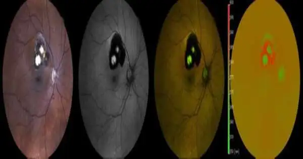
An expected 5 to 10% of visual impairment overall is brought about by the uncommon fiery eye illness uveitis. Back uveitis, in particular, is frequently associated with serious illness movement and the need for immunosuppressive treatment.In back uveitis, irritation happens in the retina and in the basic choroid that provisions it with supplements. Scientists at the Ophthalmology Department at the University of Bonn have tried variety-coded fundus autofluorescence as a strong novel indicative strategy. Fluorescence of the retina can be utilized to deduce the uveitis subtype. This is a fundamental essential for the exact finding and treatment of the illness. The outcomes have now been distributed in Scientific Reports.
Those affected by the uncommon illness back uveitis have no trouble with blurred vision, floaters, and strange light insight.Yet, the results can be serious: About five to a modest amount of visual impairment overall is brought about by uveitis. “Uveitis is an uncommon illness, yet back uveitis specifically possibly has an unfortunate occurrence and frequently requires immunosuppressive treatment,” makes sense to Dr. Maximilian Wintergerst of the Ophthalmology Department at the University of Bonn. There are various types of illness. In back uveitis, the retina or choroid in the eye becomes excited. While the retina changes light into nerve impulses over time, the choroid supplies the external layers of the retina with supplements.
Different helpful administrations
“It’s difficult to recognize the various subtypes of uveitis,” says Wintergerst. Nonetheless, since the different subtypes frequently require alternate helpful methodologies, a solid finding is even more significant. To this end, scientists from the Ophthalmology Department at the University of Bonn, along with partners from the Medical Biometry and Rheumatology Departments at the University Hospital Bonn and the University Hospital of Ophthalmology in Bern (Switzerland), explored another imaging method that can aid the finding of back uveitis.
“Our findings suggest that this ratio might be quite distinctive and useful as a marker for identifying the various posterior uveitis subtypes. It may help us to make more reliable diagnoses in the future.”
Prof. Dr. Dr. Robert Finger, co-author of the study
The group assessed variety-coded fundus autofluorescence (Spectrally Resolved Autofluorescence Imaging). The CenterVue (iCare) organization from Padua (Italy) gave the recently evolved gadget to the analysts for the assessments. This cycle includes enlightening the retina with pale blue light. The retina retains the light and refracts it again at an alternate frequency. The gadget estimates this fluorescence and divides the signs into a green and a red part.
Wintergerst makes sense of how the green-to-red proportion of the light radiated from each fiery center depends, among different elements, on the specific back uveitis subtype involved. The analysts analyzed the eyes of 45 review members. In every one of them, the specific subtype of uveitis was analyzed in advance. This included ophthalmologic assessment discoveries, lab examinations, serologic and radiologic discoveries, and at times, hereditary and interdisciplinary clinical assessments.
More robust analyses using color-coded fundus auto fluorescence
The analysts assessed the green-red proportion in the fluorescence of the fundus of the eye for around 800 fiery foci according to the patients. “Our outcomes show that this proportion can be used as a trademark and supportive as a marker for separating the different back uveitis subtypes,” states Prof. Dr. Robert Finger, co-creator of the review and head of the uveitis center at the University Hospital Bonn. “It could permit us to make more certain findings later on.” This is a major step for the Ophthalmology Department at the University of Bonn, particularly since Finger organizes the vast uveitis vault “TOFU” (Treatment Leave Choices for Non-Irresistible Uveitis, www.tofu-uveitis-register.de) along with partners from Münster. The point is to report illness movement on a drawn-out premise and to foster proposals for treatment rules.
“In the ongoing review, we present the exact specialized foundation of variety coded fundus auto fluorescence in ophthalmology as a team with our global accomplices,” explains head of division Prof. Dr. Blunt Holz. “This innovation may also enable better checking of back uveitis later on, in addition to more solid analyses.”
More information: Maximilian W. M. Wintergerst et al, Spectrally resolved autofluorescence imaging in posterior uveitis, Scientific Reports (2022). DOI: 10.1038/s41598-022-18048-4
Journal information: Scientific Reports


