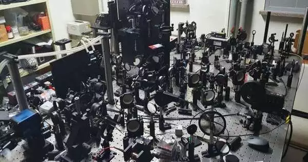
Scientists led by Partner Chief Choi Wonshik of the Foundation for Essential Science’s Center for Subatomic Spectroscopy and Elements, Teacher Kim Moonseok of The Catholic University of Korea, and Teacher Choi Myunghwan of Seoul Public University developed a new type of holographic magnifying lens.It is said that the new magnifying lens would be able to “see through” the flawless skull and be able to do high-resolution 3D imaging of the brain network inside a living mouse mind without eliminating the skull.
To examine the inner elements of a living creature utilizing light, it is important to A) convey adequate light energy to the example and B) precisely measure the signal reflected from the objective tissue. Nonetheless, in living tissues, various dispersing impacts and serious deviations will generally happen when light stirs things up around town, which makes it hard to get sharp pictures.
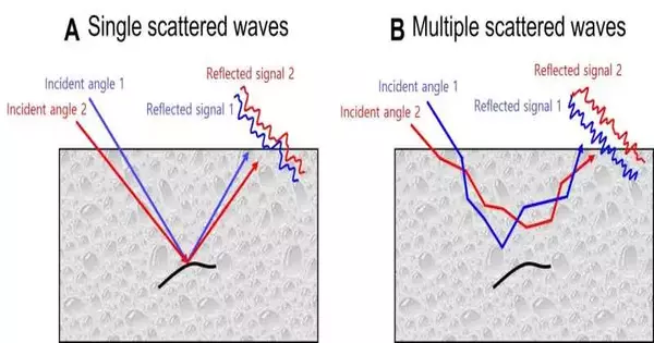
In complex designs, for example, living tissue, light goes through various dispersions, which makes the photons arbitrarily shift their course a few times as they travel through the tissue. Due to this cycle, a large part of the picture data conveyed by the light becomes destroyed. In any case, regardless of whether it is a tiny measure of mirrored light, it is feasible to notice the elements found somewhat profound inside the tissues by revising the wavefront twisting of the light that was reflected from the objective. In any case, the previously mentioned various dispersing impacts impede this remedy cycle. Hence, to get a high-goal profound tissue picture, eliminating the various dispersed waves and increasing the proportion of the single-dissipated waves is significant.

Figure 2. Qualities of the reflected sign as per the episode angle (A) Assuming the item is small or has a direct design, the waveform of the reflected sign of the single dispersed waves stays comparable in any event when the occurrence point is changed. (B) In any case, the waveform of the reflected sign of the various dispersed waves changes without likeness, even with a slight change in episode point. Utilizing these wavefront properties, single dispersing parts and various dissipating parts can be isolated from one another.
In 2019, the IBS analysts fostered a fast time-settled holographic magnifying lens that can kill various dispersions and all the while measure the adequacy and period of light. They utilized this magnifying lens to observe the brain organization of live fish without incisional medical procedure. Nonetheless, on account of a mouse, which has a thicker skull than that of a fish, it was impractical to get a brain network picture of the mind without eliminating or diminishing the skull because of serious light bending and various dispersing happening when the light goes through the bone design.
“When we originally discovered optical resonance in complex media, we received a lot of attention from academics. By integrating the efforts of bright experts in physics, life, and brain research, we have opened a new path for brain neuroimaging convergent technology, from basic concepts to practical application of seeing the neural network beneath the mouse skull.”
Professor Kim Moonseok and Dr. Jo Yonghyeon
The examination group figured out how to quantitatively dissect the connection between light and matter, which permitted them to additionally work on their past magnifying lens. In this new review, they detailed the fruitful improvement of a super-profundity, three-layered time-settled holographic magnifying lens that considers the perception of tissues to a more prominent profundity than at any other time.
In particular, the scientists conceived a strategy to choose single-dispersed waves by exploiting the way that they have comparable reflection waveforms in any event when light is input from different points. This was finished by an intricate calculation and a mathematical activity that dissects the eigenmode of a medium (a novel wave that conveys light energy into a medium), which permits the finding of a reverberation mode that boosts useful impedance (obstruction that happens when rushes of a similar stage cross-over) between wavefronts of light. This empowered the new magnifying lens to shine in excess of multiple times the light energy on the brain strands than previously, while specifically eliminating pointless signs. This permitted the proportion of single-dispersed waves versus various dissipated waves to be expanded by a few significant degrees.

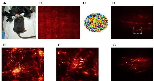
Figure 3. A brain network in the mind of a living mouse was seen without eliminating the skull. (A). The mind’s brain network was effectively imaged by involving a light source in the noticeable frequency locale. Just the skin of a living mouse was taken out, and the skull was left in one piece. (B) Utilizing the past innovation, it was unrealistic to address the perplexing variation because of the serious dispersed waves created in the skull, which makes it difficult to get any sound picture. (C) Nonetheless, the calculation created by the examination group permitted specific expulsion of various dispersing parts within the reflected sign, which allowed the wave front deviation to be revised. (D). This permitted them to determine the fine construction of brain strands inside the mind. E, F) High-resolution projection images depict osteocytes inside the mouse skull, which thrive between bone layers and dura mater, and G) a magnified brain network.
The examination group proceeded with the demonstration of this new innovation by noticing the mouse mind. Using existing technology, the magnifying lens was able to address wave front bending even at previously unthinkable depths. The new magnifying lens prevailed with regards to getting a high-goal picture of the mouse mind’s brain network under the skull. This was completely accomplished in the visible frequency without removing the mouse skull or requiring a fluorescent name.
Teacher Kim Moonseok and Dr. Jo Yonghyeon, who have fostered the groundwork of the holographic magnifying lens, said, “When we initially noticed the optical reverberation of intricate media, our work got incredible consideration from the scholarly world. From essential standards to viable use of noticing the brain network underneath the mouse skull, we have opened another way for mind neuroimaging innovation by consolidating the endeavors of gifted individuals in physical science, life, and cerebrum science. “
Partner Chief Choi Wonshik said, “For quite a while, our middle has grown in super-profundity bioimaging innovation that applies actual standards. It is normal that our current finding will enormously contribute to the improvement of biomedical interdisciplinary examination, including neuroscience and the business of accuracy metrology. “
This examination was distributed in the web-based version of the journal Science Advances on July 28.
More information: Yonghyeon Jo et al, Through-skull brain imaging in vivo at visible wavelengths via dimensionality reduction adaptive-optical microscopy, Science Advances (2022). DOI: 10.1126/sciadv.abo4366
Journal information: Science Advances


