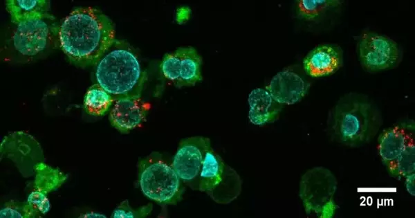
Particles known as extracellular vesicles assume a fundamental role in correspondence among cells and in numerous cell capabilities. These “membrane particles” are cargoes of specific signaling molecules, proteins, nucleic acids, and lipids that are released into the environment by cells. Sadly, only a small number of the vesicles are spontaneously formed by cells.
The items in these extracellular vesicles change contingent upon the beginning and state of the cell, as do the proteins that are moored to the vesicle surface. By analyzing extracellular vesicles isolated from blood samples, for instance, researchers use these properties to create novel methods for diagnosing cancer.
Extracellular vesicles could likewise play a critical role in the improvement of cutting-edge therapeutics. Because they come from nature, the vesicles are biocompatible and can elicit a wide range of responses in the body.
“We begin the preparation process by cultivating cancer cells and inducing cell death with chemical stressors. The cells then divide and form vesicles, which detach from the parent cell after a few hours.”
Claudio Alter, first author of the study and a doctoral student at the SNI Ph.D. School.
As a result, the researchers hope to use the particles to influence the immune system, possibly to eradicate cancer cells. As of recently, nonetheless, one significant test has been the reproducible creation of the huge amounts of homogeneous vesicles required for such examinations.
A quicker course to additional particles
Presently, a group of scientists led by Professor Jörg Huwyler from the Branch of Drug Sciences and the Swiss Nanoscience Establishment (SNI) of the College of Basel has fostered a profoundly proficient planning strategy for extracellular vesicles that conveys up to multiple times a bigger number of particles per cell and hour than traditional techniques. The new method is described in the Communications Biology journal.
“We start the readiness cycle by developing malignant growth cells, in which we prompt cell demise by adding substance stressors,” makes sense to Claudio Modify, the first writer of the review and a doctoral understudy at the SNI Ph.D. School. ” After that, the cells make vesicles, which eventually separate from the parent cell after a few hours.
With a width of 1 to 3 micrometers, these monster plasma film vesicles are unreasonably enormous for remedial applications. In the recently evolved process, they are squeezed through a channel film on various occasions to decrease their size. “After various channel passes, we get a homogeneous arrangement of nanoplasma film vesicles (nPMV) with a breadth of 120 nanometers —definitely what we want for resulting applications,” makes sense of Change.
Different origins, different uses
The researchers then compared the size, homogeneity, and protein and lipid cargo of these nPMVs to those of exosomes, which are currently the most widely used extracellular vesicles. They also looked into the nPMVs’ ability to interact with other cells. The nanoplasma membrane vesicles exhibited exosome-like characteristics in these analyses.
“Their particular freight and the presence of film-bound markers from the parent cell line offer the likelihood of involving nPMVs for remedial purposes,” says Jörg Huwyler. “At the moment, we’re mostly thinking about ways to stimulate the immune system, like in vaccines or cancer treatments with immunotherapy.”
More information: Claudio L. Alter et al, High efficiency preparation of monodisperse plasma membrane derived extracellular vesicles for therapeutic applications, Communications Biology (2023). DOI: 10.1038/s42003-023-04859-2


