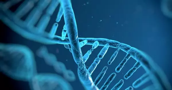
A new ‘image analysis pipeline’ is providing scientists with rapid new insights into how disease or injury have changed the body, all the way down to the individual cell level. TDAExplore combines microscopy’s detailed imaging with topology, a hot area of mathematics that provides insight into how things are arranged, and the analytical power of artificial intelligence to provide, for example, a new perspective on changes in a cell caused by ALS and where in the cell they occur, according to a cell biologist involved with the study.
Dr. Eric Vitriol, a cell biologist and neuroscientist at the Medical College of Georgia, describes a new perspective on changes in a cell caused by ALS and where in the cell they occur. It is an “accessible, powerful option” for using a personal computer to generate quantitative – measurable and thus objective – information from microscopic images, which could also be applied to other standard imaging techniques such as X-rays and PET scans, they write in the journal Patterns.
“We think this is an exciting step forward in using computers to provide us with new information about how image sets differ from one another,” Vitriol says. “What are the actual biological changes that are occurring, including those that I may not be able to see because they are too minute or because I have some sort of bias about where I should be looking?”
According to the neuroscientist, computers have our brains beat when it comes to analyzing data, not just in terms of objectivity, but also in terms of the amount of data they can assess. Computer vision, which allows computers to extract information from digital images, has been around for decades, so he and his colleague and co-corresponding author Dr. Peter Bubenik, a mathematician at the University of Florida and an expert on topological data analysis, decided to combine the detail of microscopy with the science of topology and the analytical power of AI. Topology and Bubenik were crucial, according to Vitriol.
We think this is an exciting step forward in using computers to provide us with new information about how image sets differ from one another. What are the actual biological changes that are occurring, including those that I may not be able to see because they are too minute or because I have some sort of bias about where I should be looking?
Dr. Eric Vitriol
Topology is “perfect” for image analysis, he says, because images are made up of patterns, of objects arranged in space, and topological data analysis (the TDA in TDAExplore) assists the computer in recognizing the lay of the land, in this case where actin, a protein and essential building block of the fibers, or filaments, that help give cells shape and movement, has moved or changed density. It’s an efficient system because, instead of training the computer on hundreds of images, it can learn on 20 to 25 images.
Part of the magic is that the computer is now learning the images in patches. They write that breaking down microscopy images into these pieces allows for more accurate classification, less training of the computer on what “normal” looks like, and ultimately the extraction of meaningful data.
Without a doubt, microscopy, which allows for close examination of things that are not visible to the naked eye, produces beautiful, detailed images and dynamic videos that are indispensable to many scientists. “You can’t have a medical school without sophisticated microscopy,” he says.
But, in order to understand what is normal and what happens in disease states, Vitriol requires a detailed analysis of the images, such as the number of filaments; where the filaments are in the cells, close to the edge, in the center, scattered throughout, and whether some cell regions have more. The patterns that emerge in this case tell him where actin is and how it’s organized, a major factor in its function, and where, how, and if it has changed due to disease or damage.
When he looks at the clustering of actin around the edges of a central nervous system cell, for example, he can tell that the cell is spreading out, moving around, and sending out projections that become its leading edge. In this case, the cell, which had been essentially dormant in a dish, can spread out and stretch its legs.
Some of the issues with scientists analyzing images directly and calculating what they see include the fact that it takes time and the fact that even scientists have biases. As an example, and especially with so much activity going on, their gaze may be drawn to the familiar, in Vitriol’s case, that actin at the leading edge of a cell. As he looks again at the dark frame around the cell’s periphery, clearly indicating the actin clustering there, he may conclude that this is the main point of action.
“How do I know that when I decide what’s different, it’s the most different thing, or is that just what I wanted to see?” he asks. “We want to bring computer objectivity to it, and we want to bring a higher level of pattern recognition to image analysis.”
AI is known to be capable of “classifying” things, such as recognizing a dog or a cat every time, even if the image is fuzzy, by first learning many millions of variables associated with each animal until it recognizes a dog when it sees one, but it cannot report why it is a dog. That approach, which requires so many images for training and still does not provide many image statistics, does not really work for his purposes, which is why he and his colleagues created a new classifier that is limited to topological data analysis.
The unique coupling used in TDAExplore, he says, efficiently and objectively tells scientists where and how much the perturbed cell image differs from the training, or normal, image, information that also provides new ideas and research directions.
Returning to the cell image with actin clustering along its perimeter, TDAExplore revealed that, while the “leading edge” was clearly different with perturbations, some of the most significant changes occurred inside the cell.
“A lot of my job is trying to find patterns in images that are hard to see,” Vitriol says, “because I need to identify those patterns so I can figure out how to get numbers out of those images.” His bottom lines include determining how the actin cytoskeleton, for which the filaments provide scaffolding and which in turn supports neurons, works and what goes wrong in conditions such as ALS.
According to the researchers, some machine learning models that require hundreds of images to train and classify images do not describe which part of the image contributed to the classification. Such massive amounts of data, which may include up to 20 million variables, necessitate the use of a supercomputer. Instead, the new system requires fewer high-resolution images and characterizes the “patches” that led to the chosen classification. The scientist’s standard personal computer can complete the new image analysis pipeline in a matter of minutes.
According to him, the novel approach used in TDAExplore objectively tells scientists where and how much the perturbed image differs from the training image, providing new ideas and research directions.
The ability to extract more and better information from images ultimately means that information generated by basic scientists like Vitriol, which frequently changes what is considered the facts of a disease and how it is treated, is more accurate. This could include being able to detect previously unnoticed changes, such as those identified by the new system inside the cell.
Currently, scientists use stains to improve contrast before using software to extract information about what they see in the images, such as how actin is organized into larger structures, he says. “We needed to come up with a new way to extract relevant data from images, and that is what this paper is about.”


