To notice living cells through a magnifying lens, an example is typically pressed onto a glass slide. It then, at that point, lies there tranquilly, and the cells are recognizable. The limitation is that this cutoff points how the cells act and only provides two-layered images.
Scientists from UiT The Cold College of Norway and the College Medical Clinic of North Norway (UNN) have now evolved what they are alluding to as the “cutting edge magnifying instrument.” The new innovation can take pictures of a lot bigger examples than previously, while living and working in a more regular habitat.
A significant turn of events
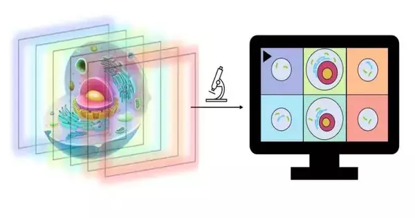
The innovation gives 3D pictures where analysts can concentrate on the littlest subtleties from a few points, obviously and noticeably arranged into various layers, all of which are in the center.
3D magnifying lenses really do exist as of now, yet they work gradually and give more unfortunate outcomes. The most widely recognized type works by keeping many pixels in series, which are then gathered into a 3D picture. This requires some investment, and frequently, they can’t deal with more than 1–5 shots per minute. It’s not unreasonable to anticipate that what you’ll photograph will be something that moves.
“We can achieve roughly 100 complete frames per second with our technology. And we believe it is possible to raise this figure. This is precisely what our prototype has demonstrated.”
Florian Ströhl, researcher at UiT.
“With our innovation, we can oversee around 100 full casings each second.” What’s more, we accept that expanding this number is conceivable. “This is exactly the very thing we have exhibited with our model,” says Florian Ströhl, a scientist at UiT.
The new magnifying lens is a supposed multifocus magnifying lens, which gives totally clear pictures arranged into various layers where you can concentrate on the cells from all points.
“It’s no joking matter. “The way that we figure out how to get this in one take, it is a tremendous turn of events,” says Ströhl.
Can see behind objects
Ströhl is correct that we are not discussing 3D in the structure that most of us are familiar with.While in a traditional 3D image, you will want to see some depth, with the new technology you will also be able to see behind objects.
Ströhl utilizes a model where you see a wilderness scene in 3D in the film.
“In an ordinary 3D picture, you can see that the timberland has a profundity, that a few leaves and trees are closer than others. You can also see the tiger hiding behind the shrubs using the same technology used in our new 3D magnifying lens.”You can see and concentrate on a few layers freely,” says Ströhl.
Currently, you don’t use a magnifying lens to look for tigers in the wild; however, for scientists, this can be a useful tool when looking for answers in the smallest details.
Concentrating on heart cells—wwhile they thump
Ströhl has worked together with scientists and specialists from the University Hospital of North Norway (UNN) in the improvement of this innovation.
In addition to other things, they work to comprehend and foster better treatment strategies for different heart illnesses.
Concentrating on a living human heart is testing, both for specialized reasons and not least for moral reasons. Hence, specialists have utilized undifferentiated organisms that are controlled so they emulate heart cells. Along these lines, they can develop natural tissue that acts as it would in a human heart, and they can review and test this tissue to see more about what’s going on.
This tissue is practically similar to a little piece of live meat, around 1 cm in size. This makes for an extremely overbearing test circumstance, where heart cells thump and are in steady movement along it in such a way that the example is too enormous to even consider examining with a conventional magnifying lens. The new magnifying instrument handles this well.
“You have this siphoning piece of meat in a bowl, which you need to take magnifying lens pictures of. You need to see the exceptionally littlest pieces of this, and you need high goals. “We have accomplished this with the new magnifying instrument,” says Ströhl.
Division 1 of the recipe
Kenneth Bowitz Larsen heads a huge lab with cutting-edge magnifying instruments that are utilized by all the exploration groups at the Workforce of Wellbeing at UiT. He has tried this new magnifying lens and is hopeful.
“The idea is brilliant; the magnifying lens they’ve built does things that business frameworks don’t,” Larsen explains.The research center he heads basically utilizes business magnifying instruments from providers like Zeiss, Nikon, and so forth.
“Then, at that point, we additionally work together with research groups like the one Florian Ströhl addresses.” “They construct magnifying lenses and test optical ideas; they are in a way like the recipe for division 1 of microscopy,” Larsen says. Larsen has extraordinary confidence in the new magnifying instrument that Ströhl has made.
The business magnifying instruments should be usable for a wide range of potential examples, while the magnifying lens Ströhl has created is more custom-fitted to a particular errand.
“It is exceptionally photosensitive, and it can portray different core interests.” It can deal with the example, and you can see both highs and lows. Furthermore, it occurs so quickly that it can be found on a continuous basis.”It’s a very quick magnifying instrument,” Larsen says.
As per Larsen, the tests up to this point show that this functions admirably, and he accepts that this kind of magnifying lens can ultimately be utilized on a wide range of tests where you check out residing things that move.
He also sees an advantage to the speed of this magnifying instrument.
“Brilliant lights are not kind to cells. “Because this magnifying instrument is so quick, it exposes the cells to a lot more limited brightening and is thus more delicate,” he understands.
The innovation is protected.
The model of the magnifying lens works and is functional. The specialists are presently chipping away at making an improved rendition that is simpler to utilize so that more individuals can work and utilize the magnifying lens.
The scientists have likewise applied for a patent and are additionally searching for modern collaborators who will form this into a magnifying lens that will be ready to move.
Meanwhile, the model will be made accessible to neighborhood accomplices who can profit from the new innovation.
“We will likewise offer it to others in Norway, assuming they have especially requested tests that they need analyzed,” says Ströhl.
The examination is distributed in Optica.
More information: Florian Ströhl et al, Multifocus microscopy with optical sectioning and high axial resolution, Optica (2022). DOI: 10.1364/OPTICA.468583
Journal information: Optica


