A group of scientists partnered with various foundations in the Republic of Korea has fostered a better approach to utilizing attractive reverberation imaging (X-ray) for harmless following of the spread of mind cues on millisecond timescales. In their paper distributed in the journal Science, the group depicts how they fostered the new innovation, its elements, and the way it worked when tried on mice. Timo van Kerkoerle and Martijn Cloos with Université Paris-Saclay and the College of Queensland, separately, have distributed a Viewpoints piece in a similar diary issue framing the work done by the group on this new effort.
An X-ray works by sending an attractive field alongside radio waves through tissue to create pictures of the tissue under study. It works on the premise that various kinds of tissue have various properties. X-ray is also used in a variety of ways, depending on the part of the body being studied.Blood-oxygen-level-subordinate practical X-ray is utilized to get pictures of the mind of a living individual—the method considers noticing changes in the blood stream in the cerebrum that act as a substitute for an expansion in neuronal activity. By and by, blood-oxygen-level-subordinate useful X-ray creates various pictures over the long haul, by and large north of a few seconds. In this new experiment, scientists have changed the manner in which an X-ray mind check is performed.
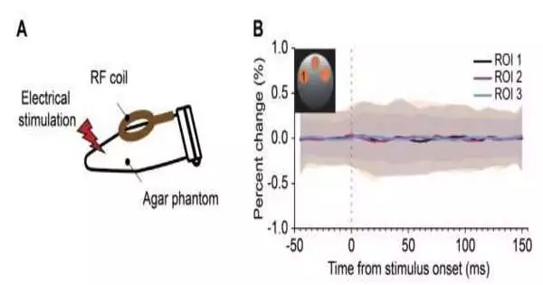
The new method, called direct imaging of neuronal action (DIANA), works by changing a customary X-ray machine to make pictures quicker — on the millisecond scale — possibly at the speed of thought. It does as such by making halfway pictures that are joined thereafter to make a high-goal picture that addresses a case of mind action. After a solitary sweep, various high-goal pictures are made that show the condition of the mind as it responds to a boost, for example, a shock of power applied to the hairs of test mice. By creating pictures so rapidly, the method permits scientists to notice the spread of mind cues, starting with one piece of the cerebrum and moving onto the next in a painless way.
The new method has so far only been tried in mice, yet the specialists are now calling it a unique advantage, proposing it could change the way that researchers concentrate on the mind — and could prompt another understanding of how it functions.
More information: Phan Tan Toi et al, In vivo direct imaging of neuronal activity at high temporospatial resolution, Science (2022). DOI: 10.1126/science.abh4340
Timo van Kerkoerle et al, Creating a window into the mind, Science (2022). DOI: 10.1126/science.ade4938
Journal information: Science


