Infections have restricted hereditary material—and scarcely any proteins—so every one of the pieces should buckle down. Zika is an extraordinary model; the infection just creates 10 proteins. Presently, in a review distributed in the journal PLOS Microorganisms, scientists at Sanford Burnham Prebys have shown how the infection accomplishes such a great deal with nearly nothing and may have distinguished a restorative weakness.
In the review, the exploration group showed that Zika’s catalyst—NS2B-NS3—is a multipurpose device with two fundamental capabilities: separating proteins (a protease) and partitioning its own twofold abandoned RNA into single strands (a helicase).
“We found that Zika’s compound complex changes capability in light of how it’s formed,” says Alexey Terskikh, Ph.D., academic partner at Sanford Burnham Prebys and senior creator of the paper. “At the point when in the shut compliance, it goes about as an exemplary protease. However, at that point, it cycles among open and super-open conformities, which permits it to snatch and afterward discharge a solitary strand of RNA—and these capabilities are fundamental for viral replication.”
“We discovered that the function of Zika’s enzyme complex changes depending on how it is shaped. When it is closed, it acts as a classic protease. However, it then cycles between open and super-open conformations, allowing it to grab and then release a single strand of RNA—functions that are critical for viral replication.”
Says Alexey Terskikh, Ph.D., associate professor at Sanford Burnham Prebys and senior author of the paper,
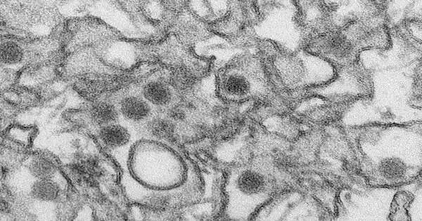
Zika is an RNA infection that is essential for a group of dangerous microorganisms called flaviviruses, which incorporate West Nile, dengue fever, yellow fever, Japanese encephalitis, and others. The infection is communicated by mosquitoes and contaminates uterine and placental cells (among other cell types), making it especially risky for pregnant women. When inside cells, the infection re-engineers them to deliver more zika.
Understanding Zika on the sub-atomic level could have a tremendous result: a remedial objective. It would be challenging to make safe medications that focus on the spaces of the chemical required for protease or helicase capabilities, as human cells have numerous comparative particles. Nonetheless, a medication that hinders Zika’s conformational changes could be successful. On the off chance that the complex can’t shape-shift, it can’t carry out its basic roles, and no new Zika particles would be created.
A productive machine
Analysts have long realized that Zika’s fundamental protein was made out of two units: NS2B-NS3pro and NS3hel. NS2B-NS3pro completes protease capabilities, cutting long polypeptides into Zika proteins. Notwithstanding, NS2B-NS3pro’s capacities to tie single-abandoned RNA and assist with isolating the twofold abandoned RNA during viral replication were as of late found.
In this review, the analysts rested on late precious stone designs and utilized protein natural chemistry, fluorescence polarization, and PC demonstrating to analyze NS2B-NS3pro’s life cycle. NS3pro is associated with NS3hel (the helicase) by a short amino corrosive linker and becomes dynamic when the complex is in its shut compliance, similar to a shut accordion. The RNA restriction happens when the complex is open, while the complex should progress through the super-open compliance to deliver RNA.
These conformational changes are driven by the elements of NS3hel movement, which broadens the linker and ultimately “yanks” the NS3pro to deliver RNA. NS3pro is secured to within the host cell’s endoplasmic reticulum (trama center)—a key organelle that helps shepherd cell proteins to their suitable objections—through NS2B and, while in the shut compliance, cuts up the Zika polypeptide, producing every single viral protein.
On the opposite side of the linker, NS3hel isolates Zika’s twofold abandoned RNA and helpfully hands a strand over to NS3pro, which has decidedly charged “forks” to take hold of the adversely charged RNA.
“There’s an exceptionally pleasant notch of positive charges,” says Terskikh. “Along these lines, RNA just normally follows that notch. Then the intricate movements to the shut compliance and delivery of the RNA.”
As NS3hel comes forward to snatch the twofold abandoned RNA, it pulls the complex with it; nonetheless, since the NS3pro is moored in the trama center film and the linker can broaden up until this point, the complicated snaps into the super-open conformity and delivers RNA. The complicated then loosens up back to the open conformity, prepared for another cycle.
In the mean time, when NS3pro distinguishes a viral polypeptide to cut, it powers the complex into shut compliance, turning into a protease. The creators refer to this interaction as “invert inchworm,” in light of the fact that snatching and delivering the single-abandoned RNA looks like inchworm developments, but in reverse, with the jaws (the protease) limping along.
As well as giving a potential remedial objective to Zika, this nitty-gritty comprehension could be applied to other flaviviruses that share comparative sub-atomic hardware.
“Forms of the NS2B-NS3pro complex are found all through the flaviviruses,” says Terskikh. “It might actually comprise a totally different class of medication focused on numerous infections.”
More information: Sergey A. Shiryaev et al. Dual Function of Zika Virus NS2B-NS3 Protease, PLOS Pathogens (2023). DOI: 10.1371/journal.ppat.1011795


