Light scattering happens when light interacts with different objects or materials, causing it to alter direction and intensity. This scattering can tell a great deal about the qualities and structures of the objects with which the light interacts, including biological specimens.
A new approach for reconstructing the 3D refractive index distribution of biological samples with several forms of light scattering has been created by researchers. The algorithm aids in the optimization of a new imaging technique known as intensity diffraction tomography (IDT).
This discovery will be presented at the Optica Imaging Congress by Jiabei Zhu of Boston University. The hybrid meeting will take place in Boston, Massachusetts, from August 14 to August 17, 2023.
“3D quantitative phase imaging (QPI) has superior features for a variety of biomedical imaging applications.” QPI, as a label-free technology, may photograph transparent living organisms and cells without the use of exogenous contrast ants or dyes, which can cause phototoxicity and damage to the sample,” explains Zhu.
Compared with traditional phase-contrast and differential interference contrast microscopy, QPI not only provides high-contrast morphological information but gives quantitative phase information as well. Specifically, 3D QPI can provide high-resolution 3D refractive index (RI) distribution inside the samples
Jiabei Zhu
“Compared with traditional phase-contrast and differential interference contrast microscopy, QPI not only provides high-contrast morphological information but gives quantitative phase information as well. Specifically, 3D QPI can provide high-resolution 3D refractive index (RI) distribution inside the samples. This valuable information can facilitate the research on hematology, neurology, and immunology, helping the diagnosis of disease and infection.”
Although 3D imaging techniques can be utilized to analyze dense biological samples, it is difficult to achieve both high-speed acquisition and good resolution. Label-free phase tomography techniques such as IDT help to circumvent this constraint. They can be carried out with the help of a programmable LED array, which can be easily fitted to a regular microscope.
Zhu’s team recently developed two IDT approaches, annular IDT (aIDT) and multiplexed IDT (mIDT), that accelerate picture capture enough to observe dynamic biological material. Annular IDT (aIDT) use an LED ring that matches the numerical aperture of the objective, whereas multiplexed IDT (mIDT) employs multiple LEDs to illuminate the sample at the same time.
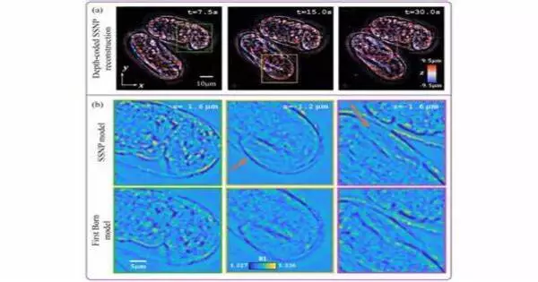
The researchers opted to create a new method after discovering that existing IDT reconstruction algorithms did not perform well with their novel techniques due to the usage of high-NA objectives. It employs a multiple scattering model based on the split-step non-paraxial (SSNP) approach, which was developed lately to overcome constraints in optical diffraction tomography.
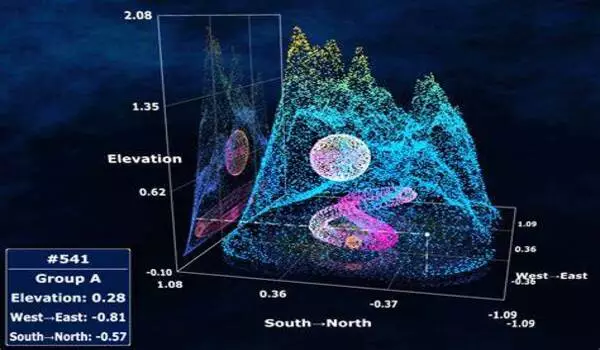
The researchers demonstrated that employing aIDT to apply the new IDT reconstruction method to buccal epithelial cells allowed for easy discrimination of cells at different depths, restoration of cell borders and membranes, and observation of natural bacteria around the cells. They also used mIDT on a thick multi-scattering live C. elegans embryo. The reconstructed images revealed details about how the worms were folded, and the single-depth cross-section revealed morphological features like the cells’ contour, the buccal cavity, and the worm’s tail.
Overall, the experiments demonstrated that the researchers were able to achieve high-quality images with a wide field of view by extending the SSNP approach to IDT.