A research team at Peking University in China has created a small three-photon microscope that has been successful in capturing deep images of freely moving mice’s brains, marking a new advancement in multiphoton microscopy.
The microscope, which only weighed 2.17 grams, managed to stabilize imaging of the brain cortex and hippocampal neurons of freely moving mice, an innovation that shows promise as a way to unravel the mysteries of the human brain.
The investigation was reported in Nature Methods.
What does the research concern?
Global brain research has focused on mapping the connectivity and functional dynamics of the human brain, which has trillions of synapses and billions of neurons. One of the research tools is wearable microscopic imaging equipment that can be used on small animals.
The National Biomedical Imaging Center at Peking University is directed by Cheng Heping, who leads a team that has been working on creating such devices for years.
In 2017, the team created the world’s smallest two-photon microscope, which weighs only 2 points 2 grams. They used it to capture live images of mouse cerebral cortex synaptic and neuronal activity while the mice were moving freely.
After four years of work, a more advanced two-photon microscope increased the imaging field by seven and a half times and was able to record three-dimensional images of the functional signals of cerebral cortex neurons, according to Cheng, a fellow academician of the Chinese Academy of Sciences.
Image stacks from the PPC and dorsal hippocampal CA1 of a mouse with its head fixed were reconstructed in three dimensions. a stack with 2-m intervals, starting at the cortical surface and going through PPC, CC, and CA1’s SP layer’s bottom. Signals from the third harmonic generation are magenta; green, GCaMP6s-labeled neurons. 250 x 250 m2 is the FOV. On the top left corner, the imaging depth is visible. Credit: Nature Methods (2023). DOI: 10.1038/s41592-023-01777-3.
How is the new technology superior?
Compared to earlier miniature multiphoton microscopes, the new three-photon microscope has a greater imaging depth. It was created by a team led by Cheng and Wang Aimin, a researcher at Peking University.
It penetrated the entire cortex and callosum of freely moving mice and captured images of calcium activity in the hippocampal region at a depth of up to 1.2 millimeters, which had been an enormous challenge for neuroscientists all over the world.
Combining fluorescent molecules with calcium indicator activity, which reflects the cellular activity of neurons, allows for monitoring. Fluorescence has a limited penetration distance in brain tissue due to light scattering, particularly in the callosum beneath the cortex, which restricts the depth of the imaging.
Cortex and callosum sit above the hippocampus. According to Zhao Chunzhu, a team member from the College of Future Technology at Peking University, previous small multiphoton microscopes around the globe had been unable to record their non-invasive imaging.
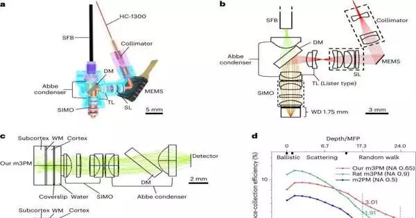
A novel optical setup that maximized the efficiency of scattering fluorescence’s collection allowed the team’s three-photon microscope to successfully image the deep brains of mice that were moving around freely.
In addition, the study found that the microscope has low phototoxicity, which prevents any visible photobleaching or photodamage during the prolonged observation of neuronal activities.
More information: Chunzhu Zhao et al, Miniature three-photon microscopy maximized for scattered fluorescence collection, Nature Methods (2023). DOI: 10.1038/s41592-023-01777-3


