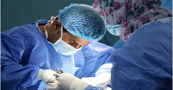
At the point when a patient goes through a careful activity to eliminate a growth or treat an illness, the course of a medical procedure is frequently not foreordained. To conclude how much tissue should be taken out, specialists should find out about the condition they are treating, including a growth’s edges, its stage and whether a sore is harmful or harmless — judgments that frequently pivot after gathering, examining, and diagnosing an illness while the patient is on the surgical table.
At the point when specialists send tests to a pathologist for assessment, both speed and precision are of the pith. The ongoing best quality level methodology for inspecting tissues frequently takes too lengthy and a quicker approach, which includes freezing tissue, can present relics that can muddle diagnostics.
Another concentrate by examiners from the Mahmood Lab at the Brigham and Ladies’ Clinic, an establishing individual from the Mass General Brigham medical care framework, and partners from Bogazici College fostered a superior way; the strategy use man-made reasoning to decipher between frozen segments and the best quality level methodology, working on the nature of pictures to build the precision of fast diagnostics. Discoveries are distributed in Nature Biomedical Designing.
“We are utilizing the potential of artificial intelligence to address an age-old challenge at the junction of surgery and pathology. Making a quick diagnosis from frozen tissue samples is difficult and needs specific knowledge, but it is a key step in caring for patients during surgery.”
Faisal Mahmood, Ph.D., of the Division of Computational Pathology at BWH.
“We are utilizing the force of man-made reasoning to resolve a deep rooted issue at the convergence of medical procedure and pathology,” said relating creator Faisal Mahmood, Ph.D., of the Division of Computational Pathology at BWH. “Making a fast finding from frozen tissue tests is testing and requires specific preparation, yet this sort of conclusion is a basic move toward really focusing on patients during medical procedure.”
For making last findings, pathologists use formalin-fixed and paraffin-implanted (FFPE) tissue tests — this strategy jam tissue such that produces great pictures yet the cycle is arduous and normally requires 12 to 48 hours. For a quick finding, pathologists utilize a methodology known as cryosectioning that includes quick freezing tissue, cutting segments, and noticing these slim cuts under a magnifying lens. Cryosectioning requires minutes instead of hours yet can twist cell subtleties and split the difference or tear fragile tissue.
Mahmood and co-creators fostered a profound learning model that can be utilized to decipher between frozen segments and more usually utilized FFPE tissue. In their paper, the group showed the way that the strategy could be utilized to subtype various types of disease, including glioma and non-little cell cellular breakdown in the lungs.
The group approved their discoveries by selecting pathologists to a peruser concentrate on in which they were approached to make a finding from pictures that had gone through the man-made intelligence strategy and customary cryosectioning pictures. The man-made intelligence strategy further developed picture quality as well as worked on analytic exactness among specialists. The calculation was likewise tried on freely gathered information from Turkey.
The creators note that later on, planned clinical examinations ought to be led to approve the man-made intelligence strategy and decide whether it can add to analytic exactness and careful dynamic in genuine clinic settings.
“Our work shows that man-made intelligence can possibly make a period delicate, basic finding simpler and more open to pathologists,” said Mahmood. “Also, it might actually be applied to a disease medical procedure. It opens up numerous opportunities for further developing finding and patient consideration.”
More information: A deep-learning model for transforming the style of tissue images from cryosectioned to formalin-fixed and paraffin-embedded, Nature Biomedical Engineering (2022). DOI: 10.1038/s41551-022-00952-9


