Exploring how tumors develop and spread has routinely been done on two-layered, level societies of cells, which is altogether different to the three-layered design of cells in the body. 3D cell societies that integrate tissue material have been grown, yet the techniques to gauge how disease cells use power to spread have been deficient.
Presently, scientists have fostered another strategy for 3D culture to precisely measure how disease cells create the power to spread inside tissue. “We have applied the strategy for examination of early movement of bosom disease,” says Juho Pokki, a key examiner at Aalto University who drove the exploration.
This review, a cooperation between researchers at Aalto University and Stanford University, was published in the journal Nano Letters.
Nanospheres measure force beats that amass into more grounded powers.
An essential growth can take shape inside the bosom’s mammary pipe, where the harmful cells are bound by a unique film, called a cellar layer. Bosom disease cells are bigger than the pores in these films, so they should get through to spread to different tissues. Already, analysts feel that cells use proteins to break up films, but presently it is perceived that bosom disease cells utilize one more system, including cell bulges, to go through the layers.
“Breast cancer cells employ the forces created by the protrusions to open up channels inside the membrane material in this method. The cancer cells then infiltrate the surrounding tissue and may migrate to blood vessels to spread throughout the body. In reality, a basement membrane surrounds the blood vessels. Breast cancer cells may employ a similar technique to break through basement membranes.”
Juho Pokki, a principal investigator at Aalto University
“In this system, bosom disease cells use powers created by the bulges to open up channels inside the film material.” Then, the disease cells enter the encompassing tissue and may go further into the veins to spread to the remainder of the body. Truth be told, the veins are likewise encircled by a cellar film. Bosom disease cells possibly utilize a comparable system to get through into those cellar films, “which makes sense of Pokki.” “Teacher Ovijit Chaudhuri’s gathering at Stanford initially tracked down this bulge system in 2018. Cooperation with his gathering has been the key to the physiological meaning of this work, “says Pokki.

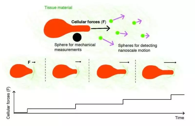
Another method estimates the power created by disease cells with a natural magnifying lens that identifies biocompatible circles inside the tissue material. Cell powers are calculated using data from two types of circles: one that recognizes nanoscale movement and another that acts on mechanical properties.The method demonstrates that disease cells generate abilities in a stepwise manner, and the abilities accumulate within the tissue material, encompassing a bosom malignant growth cancer.Credit: Juho Pokki/Aalto University.
The new review utilizes 3D cell societies made out of bosom disease cells and standard cellar film material. Inside the 3D societies, analysts inserted two sorts of biocompatible circles: one sort moved alongside powers created by disease cells, and the other kind estimated force-obliging mechanics. A modified fluorescence magnifying lens was used to record and track these circles at nanoscales.
This permitted the analysts to gauge the power beats coming from disease cells. “Past examinations had estimated movement of cell bulges over longer timeframes, yet our review demonstrated the way that a ton can occur in only 15 minutes. We saw nanoscale development and power beats inside a couple of moments, which is surprising. Further, these heartbeats amass, bringing about more grounded powers applied to the film material, “says Pokki.
“This is right now the most reliable strategy for estimating how cell powers are created in 3D culture,” adds Pokki.
Toward a more effective and customized drug advancement
Bosom disease is the most well-known type of malignant growth for women worldwide. Consistently, in excess of 350,000 women are determined to have bosom disease in the European Union alone.
Developing medicine against breast cancer is costly, time-consuming, and frequently wasteful because less than 5% of medicine candidates chosen using 2D cell societies and creature tests are viable in human clinical trials.
“Our strategies give more precise computational information on cell powers during attack by bosom disease cells.” Our gathering combines strategies with microscopy innovation to make 3D cell culture tests more reproducible. I accept that mechanical advancements will ultimately help pre-clinical examination. We have previously begun a connected task in the space of customized disease medication, “understands Pokki.
More information: Luka Sikic et al, Nanoscale Tracking Combined with Cell-Scale Microrheology Reveals Stepwise Increases in Force Generated by Cancer Cell Protrusions, Nano Letters (2022). DOI: 10.1021/acs.nanolett.2c01327
Journal information: Nano Letters


