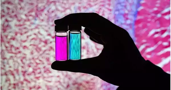
New imaging agents that can simultaneously illuminate multiple biomarkers will soon allow cancer surgeons to have a more comprehensive view of tumors during surgery, according to a report from the University of Illinois at Urbana-Champaign. The fluorescent nanoparticles, enveloped by the layers of red platelets, target growths better than current clinically supported colors and can produce two particular signs because of only one light emission, an element that could be useful to specialists to recognize growth borders and distinguish metastatic diseases.
The group’s paper, “Cell-film-covered nanoparticles for growth outline and subjective assessment of disease biomarkers at single frequency excitation in murine and apparition models,” is distributed in ACS Nano.
According to research group leader Viktor Gruev, an Illinois professor of electrical and computer engineering, the imaging agents can be used in conjunction with bioinspired cameras, which the researchers previously developed for real-time surgical diagnosis. In the new study, the researchers demonstrated their brand-new dual-signal nanoparticles in live mice and tumor phantoms, which are three-dimensional models that resemble tumors and their surroundings.
“Imaging one biomarker is insufficient for detecting all cancer. Some tumors may be missed. When a second or third biomarker is added, the likelihood of eradicating all cancer cells increases, as does the possibility of a better prognosis for the patients.”
Group leader Viktor Gruev, an Illinois professor of electrical and computer engineering.
“Imaging just one biomarker is not enough to find all the cancer,” says the author. It might overlook some tumors. In the event that you present a second or a third biomarker, the probability of eliminating all disease cells increments, and the probability of an improved result for the patient increments,” said Gruev, who likewise is a teacher in the Carle Illinois School of Medication. “Our group is leading the way with multiple-targeted drugs and imaging agents because we have camera technology that can simultaneously image multiple signals.”
According to Illinois postdoctoral researcher Indrajit Srivastava, the paper’s first author, the traditional procedure involves removing a tumor and sending it to a pathologist for evaluation, which can take anywhere from hours to days. As exploration has pushed toward ongoing diagnostics, a few difficulties have forestalled wide application: According to Srivastava, many tumor-targeted imaging agents only reach a small portion of their tumor targets and are quickly eliminated from the bloodstream before accumulating in the liver.
“A couple of individuals before us have utilized nanoparticles covered with red platelets and found they circle longer — a couple of days. The same thing was observed in our mice: the layer-covered nanoparticles circled longer in the blood, with diminished take-up in the liver. Since they were flowing longer, a greater amount of the imaging specialists collected in the cancers, giving us a more grounded fluorescent sign,” Srivastava said.
One biomarker that is prevalent in early cancer and one that is prevalent in late-stage cancer, which is more likely to be metastatic, are the two biomarkers that are the targets of the new imaging agents. The probes successfully distinguished cancerous tissue from healthy tissue and the two signals from one another, the researchers discovered.
“This is appealing for use in surgery because it might help determine precisely where to cut. Multiple signals provide a more comprehensive picture of the tumor. Additionally, it might inform a surgeon that “this may be metastatic, and you may want to be more aggressive in your removal.” Srivastava said.
Another advantage for surgical applications is that instrumentation is much smaller when only one wavelength of laser light is required to elicit multiple signals as opposed to multiple lasers for each required wavelength, according to Gruev.
The specialists intend to foster more growth imaging specialists that focus on various markers and to push ahead with additional preclinical and clinical investigations utilizing their double-sign colors with the careful goggles they have created.
Gruev stated, “We need investments both in the imaging camera technology and in the tumor-targeting agents in this battle to ensure we remove all cancer cells during surgery.” As we get closer and closer to clinical trials, this work is helping us better understand and direct our holistic approach.
More information: Indrajit Srivastava et al, Cell-Membrane Coated Nanoparticles for Tumor Delineation and Qualitative Estimation of Cancer Biomarkers at Single Wavelength Excitation in Murine and Phantom Models, ACS Nano (2023). DOI: 10.1021/acsnano.3c00578


