The team from the University of Science and Technology of China of the Chinese Academy of Sciences, led by Prof. Li Chuanfeng and Dr. Xu Jinshi, made progress in enhancing the fluorescence of single silicon carbide spin defects in a study that was published online in Nano Letters.
The scientists utilized surface plasmons to notably improve the fluorescence splendor of single silicon carbide twofold opening PL6 variety focuses, prompting an improvement in the effectiveness of twist control utilizing the properties of co-planar waveguides. The coherence properties of the color centers are not compromised by this low-cost method, nor does it necessitate complex micro-nano processing technology.
The brightness of the fluorescence of spin color centers in solid-state systems is an important parameter for real-world quantum applications. Spin color centers are essential for processing quantum information.
The fabrication of solid immersion lenses, nanopillars, bull’s eye structures, photonic crystal microcavities, and fiber cavities are all common methods for coupling spin color centers with solid-state micro-nanostructures, which are traditionally used to boost fluorescence. However, there are still obstacles, such as the difficulty of aligning particular color centers with micro-nano structures and the susceptibility of color center spin properties to complex micro-nano fabrication processes.
The team pioneered a novel strategy by utilizing plasmons to enhance the fluorescence of silicon carbide spin centers. Chemical and mechanical polishing were used by the researchers to create a thin film of silicon carbide with a thickness of about 10 micrometers. Near-surface divacancy color centers were created in the film by employing ion implantation technology.
Van der Waals forces were used to adhere the flipped film to a coplanar gold waveguide-coated silicon wafer. The near-surface color centers were able to be influenced by the gold waveguide’s surface plasmons thanks to this positioning, which increased their fluorescence.
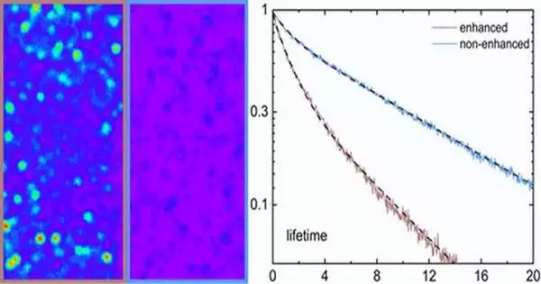
The researchers were able to make a single PL6 color center seven times brighter using an objective lens with a numerical aperture of 0.85 and the enhancement effect of surface plasmons. With an oil focal point with a mathematical gap of 1.3, the fluorescence of the variety place surpassed 1,000,000 counts per second.
In addition, by adjusting the film thickness using a reactive ion etching process, the researchers were able to precisely alter the distance between the near-surface color center and the coplanar waveguide, enabling them to investigate the optimal operating range. In addition to producing surface plasmons, the coplanar gold waveguide is capable of effectively radiating microwaves, significantly increasing spin control’s effectiveness.
The coplanar waveguide expanded the Rabi recurrence of a solitary PL6 variety focus by multiple times under a similar microwave power contrasted with that in ordinary microwave radiation techniques.
Besides, the scientists explored the component of fluorescence improvement. They confirmed that surface plasmons enhanced fluorescence brightness by increasing the radiative transition rate of the color center energy level by fitting the autocorrelation function with a three-level model and measuring the non-resonant excitation fluorescence lifetime.
They also discovered that the fluorescence brightness of the color center decreased as the interaction distance decreased due to the quenching effect of surface plasmons.
In silicon carbide films, this is the first application of plasmon-enhanced fluorescence from near-surface spin color centers. The arrangement of the coplanar gold waveguide is direct, without complex improvement designs or arrangement processes. A significant advancement in the application of silicon carbide materials to the field of quantum science is the fact that this method also enhances the fluorescence of other spin color centers in silicon carbide.
More information: Ji-Yang Zhou et al, Plasmonic-Enhanced Bright Single Spin Defects in Silicon Carbide Membranes, Nano Letters (2023). DOI: 10.1021/acs.nanolett.3c00568


