A group of designers and neuroscientists has shown interestingly that human mind organoids embedded in mice have laid out a useful network for the creatures’ cortex and answered outer tactile cues. The embedded organoids responded to visual boosts similarly as encompassing tissues, a perception that scientists had the option to make continuously for a while thanks to a creative trial arrangement that joins straightforward graphene microelectrode clusters and two-photon imaging.
The group, led by Duygu Kuzum, an employee in the College of California San Diego Branch of Electrical and PC Designing, subtleties their discoveries in the Dec. 26 issue of the journal Nature Interchanges. Kuzum’s group teamed up with analysts from Anna Devor’s lab at Boston College, Alysson R. Muotri’s lab at UC San Diego, and Fred H. Gage’s lab at the Salk Foundation.
Human cortical organoids are gotten from human incited pluripotent immature microorganisms, which are normally gotten from skin cells. These mind organoids have recently emerged as promising models for focusing on the improvement of the human cerebrum as well as a variety of neurological conditions.
“No other investigation has been able to simultaneously record optically and electrically. Our findings show that visual stimuli elicit electrophysiological responses in the organoids that correspond to the responses in the surrounding cortex.”
Madison Wilson, the paper’s first author and a Ph.D. student in Kuzum’s research group at UC San Diego.
Yet, as of recently, no examination group had the option to show that human mind organoids embedded in the mouse cortex had the option to have similar useful properties and respond to boosts similarly. This is because the technologies used to record mind capability are limited and, in general, unsuitable for recording actions that last only a few milliseconds.
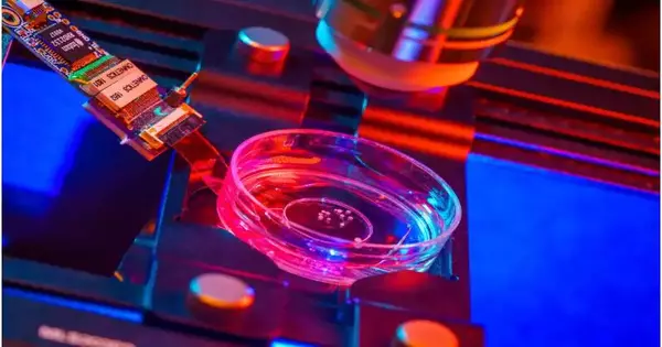
The UC San Diego-Drove team could address this issue by developing tests that connect microelectrode clusters made of simple graphene and two-photon imaging, a microscopy method that can image living tissue as thin as one millimeter.
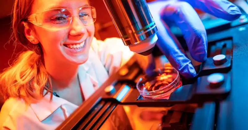
Madison Wilson, a PhD understudy at UC San Diego, is the first author of the review showing that human mind organoids embedded in mice have laid out a useful network for the creatures’ cortex and answered outer tactile boosts.
“No other review has had the option to record optically and electrically simultaneously,” said Madison Wilson, the paper’s most memorable creator and a Ph.D. understudy in Kuzum’s exploration bunch at UC San Diego. “Our tests uncover that visual boosts summon electrophysiological reactions in the organoids, matching the reactions from the encompassing cortex.”
The scientists trust that this mix of creative brain recording innovations to study organoids will act as a novel stage to completely assess organoids as models for mental health and illness and examine their utilization as brain prosthetics to reestablish capability in lost, declined, or harmed mind locales.
“This trial arrangement opens up uncommon doors for examinations of human brain network-level dysfunctions and basic formative mind illnesses,” said Kuzum.
Kuzum’s lab previously cultivated the straightforward graphene anodes in 2014 and has been propelling the innovation from that point forward. The analysts utilized platinum nanoparticles to bring down the impedance of graphene anodes by multiple times while keeping them straightforward. The low-impedance graphene anodes can record and picture neuronal action at both the macroscale and single-cell levels.
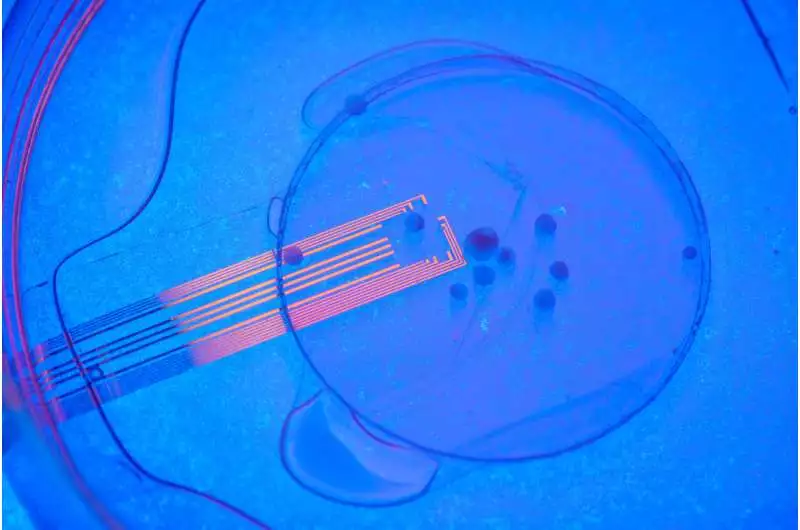
By placing a number of these cathodes on top of the relocated organoids, scientists were able to continuously record brain activity electrically from both the embedded organoid and the surrounding host cortex.They also discovered that mouse veins developed into the organoid, supplying vital nutrients and oxygen to the embed using two-photon imaging.
A visual boost was applied by scientists using an optical white light.prodded the mice with embedded organoids while the mice were under two-photon microscopy. They noticed electrical action in the anode channels over the organoids, showing that the organoids were responding to the boost similarly to the encompassing tissue. Through useful associations, the electrical action spread from the area closest to the visual cortex in the embedded organoids region.
The analysts noticed electrical action in the anode channels over the organoids, showing that the organoids were responding to the boost similarly to the encompassing tissue.
Also, their low-clamor, straightforward graphene anode innovation empowered the electrical recording of spiking action from the organoid and the encompassing mouse cortex. Graphene accounts showed expansions in the force of gamma motions and stage locking of spikes from organoids to slow motions from mouse visual cortex. These discoveries propose that the organoids had laid out synaptic associations with encompassing cortex tissue three weeks after implantation and got useful contributions from the mouse mind. The researchers ran these ongoing multimodal tests for a long time and discovered a useful and morphological mix of embedded human mind organoids with the host mouse cortex.
The following stages incorporate longer tests, including neurological illness models as well as calcium imaging in the trial set up to picture spiking action in organoid neurons. Different strategies could likewise be utilized to follow axonal projections among organoids and mouse cortex.
“That is what we anticipate, in the future, this combination of undeveloped cells and neurorecording advances will be used for demonstrating illness under physiological conditions, analyzing applicant medicines on quiet, unambiguous organoids, and assessing organoids’ capacity to reestablish explicit lost, declined, or harmed mind areas,” Kuzum said.
More information: Madison N. Wilson et al, Multimodal monitoring of human cortical organoids implanted in mice reveal functional connection with visual cortex, Nature Communications (2022). DOI: 10.1038/s41467-022-35536-3
Journal information: Nature Communications


