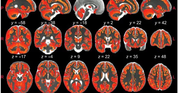
Dr. Silvia de Santis and Dr. Santiago Canals of the Institute of Neurosciences UMH-CSIC (Alicante, Spain) have used dispersion-weighted Magnetic Resonance Imaging to image interestingly and exhaustively cerebrum aggravation.This nitty-gritty “X-beam” of aggravation can’t be acquired with customary MRI, yet requires information-obtaining strategies and extraordinary numerical models. When the technique was created, the analysts had the option to measure the modifications in the morphology of the different cell populations engaged in the fiery cycle in the cerebrum.
A creative technique created by the scientists has made conceivable this significant forward leap, which is distributed today in the journal Science Advances and which might be urgent to steer the review and treatment of neurodegenerative illnesses.
“This is the first time that the signal from this sort of MRI (dw-MRI) has been proven to detect microglial and astrocyte activation, with distinct footprints for each cell population. This method represents post-mortem morphological changes substantiated by quantitative immunohistochemistry,”
Dr. Silvia de Santis and Dr. Santiago Canals, both from the Institute of Neurosciences UMH-CSIC
The exploration demonstrates that dissemination weighted MRI can harmlessly and differentially recognize the enactment of microglia and astrocytes, two sorts of synapses that are at the premise of neuroinflammation and its movement.
Degenerative cerebrum illnesses, for example, Alzheimer’s and different dementias, Parkinson’s or numerous sclerosis, are a squeezing and troublesome issue to address. Supported actuation of two sorts of synapses, microglia and astrocytes, prompts ongoing irritation in the mind that is one of the reasons for neurodegeneration and adds to its movement.
There is an absence of painless methodologies prepared to explicitly portray mind irritation in vivo. The momentum highest quality level is positron outflow tomography (PET), but it is hard to summarize and is related to openness to ionizing radiation, so its utilization is restricted to weak populations and in longitudinal examinations, which require the utilization of PET more than once over a period of years, just like the case in neurodegenerative sicknesses.
One more downside of PET is its low spatial goal, which makes it unacceptable for imaging small designs with the additional disadvantage that irritation explicit radiotracers are communicated in numerous cell types (microglia, astrocytes, and endothelium), making it difficult to separate between them.
Dissemination weighted MRI has the unique ability to image mind microstructure in vivo painlessly and with high resolution by capturing the irregular development of water particles in the cerebrum parenchyma to produce contrast in MRI images.
Imaginative procedure
In this review, specialists from the UMH-CSIC Neurosciences Institute have fostered a creative technique that permits imaging of microglial and astrocyte enactment in the dark matter of the cerebrum utilizing dissemination weighted attractive reverberation imaging (dw-MRI).
It has been shown that the signal from this sort of MRI (dw-MRI) can identify microglial and astrocyte initiation, with explicit impressions for every cell populace. The methodology we have utilized mirrors the morphological changes approved after death by quantitative immunohistochemistry, “the analysts note.
They have additionally shown that this method is delicate and explicit for identifying irritation with and without neurodegeneration, so the two circumstances can be separated. Moreover, it makes it conceivable to separate between irritation and demyelination, which is normal for numerous sclerosis.
Silvia de Santis says this work likewise shows the translational worth of the methodology utilized in a partner of sound people at high goal “in which we played out a reproducibility examination. The huge relationship with known microglia thickness designs in the human cerebrum upholds the handiness of the technique for creating solid glia biomarkers. We accept that describing, utilizing this strategy, significant parts of tissue microstructure during irritation, painlessly and longitudinally, can colossally affect how we might interpret the pathophysiology of many mind conditions and can change flow analytic practice and treatment observing techniques for neurodegenerative infections. “
To approve the model, the specialists involved a laid out worldview of irritation in rodents in light of the intracerebral organization of lipopolysaccharide (LPS). In this worldview, neuronal feasibility and morphology are protected while prompting, in an initial enactment of microglia (the cerebrum’s resistant framework cells), and in a delayed way, an astrocyte reaction. This worldly succession of cell events allows glial reactions to be briefly separated from neuronal degeneration and the mark of receptive microglia studied independently of astrogliosis.
To confine the engraving of astrocyte initiation, the specialists rehashed the examination by pretreating the creatures with an inhibitor that briefly removes around 90% of microglia. Accordingly, utilizing a laid out worldview of neuronal harm, they tried whether the model had the option to disentangle neuroinflammatory “impressions” with and without attending to neurodegeneration. “This is basic to exhibit the utility of our methodology as a stage for the disclosure of biomarkers of fiery status in neurodegenerative illnesses, where both glia enactment and neuronal harm are central participants,” they write.
At long last, the scientists utilized a laid out worldview of demyelination, in view of the central organization of lysolecithin, to show that the biomarkers created don’t mirror the tissue modifications much of the time found in cerebrum problems.
