A hallucinogenic film of pushed cells under a magnifying lens sent a group of kidney physiologists and scholars from the College of Pittsburgh and Carnegie Mellon College on an excursion to settle a secret: how do cells control their volume?
Their review, distributed today in Cell, makes sense of how the researchers utilized a tad of luck to draw an obvious conclusion to a riddle originally proposed thirty years prior.
“We were doing live fluorescence imaging tests that were irrelevant to this review, and when we added a salt solution for the cells, the inner cytoplasmic material quickly transformed into a fluorescent astro light,” said Daniel Shiwarski, Ph.D., a postdoctoral examination individual at Carnegie Mellon College, depicting how he and his better half, co-lead creator Cary Boyd-Shiwarski, M.D., Ph.D., transformed a happy piece of trial and error into a startling finding.
“Cytosol is found inside cells, and most people believe that this cytosol is diffuse, with all kinds of molecules floating around in a nicely mixed solution.”
Arohan R. Subramanya, M.D., associate professor in the Renal-Electrolyte Division
“I took a gander at her, and she asked me what was happening, similar to I should be aware,” he said. “Also, I said, ‘I have no clue, yet I believe it’s likely something significant!'”
Researchers at the College of Pittsburgh and Carnegie Mellon College tackled a decades-old secret in regards to how cells control their volume. In this video, WNK kinases (a sort of protein) are fluorescent and diffuse all through the cell. When presented with a salt arrangement, they mix into bigger beads, seeming to be the radiant green goo in an astrolight. This cycle, called “stage division,” is the way the phone realizes it needs to bring both water and particles back in, getting back to its unique state in no time. Credit: Boyd-Shiwarski, et al., Cell (2022).
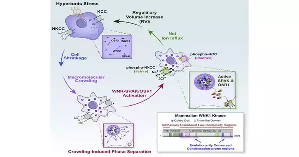
When cells are presented with an unexpected external stressor, for example, elevated degrees of salt or sugar, their volume can diminish. In the mid 1990s, researchers suggested that phones reestablish their volume by some way or another by checking their protein focus, or how “swarmed” it was inside the cell. Yet, they didn’t have any idea how the cell detected congestion.
Then, in the mid 2000s, a kind of protein called With-No-Lysine kinases, or “WNKs,” were found. For quite a long time, researchers thought that WNK kinases were switching cell shrinkage, yet the way in which they did this was likewise a secret.
The new examination tackles the two riddles, divulging how WNK kinases enact the “switch” that profits cell volume to harmony through a cycle called stage division.
“A cell contains cytosol, and most people perceive this cytosol to be diffuse, with a wide range of particles drifting around in an impeccably blended arrangement,” said senior writer Arohan R. Subramanya, M.D., academic partner in Pitt’s Institute of Medication and staff doctor at the VA Pittsburgh Medical Care Framework.
Yet, there has been this change in outlook in our reasoning about how cytosol functions. It’s truly similar to an emulsion with a lot of nearly nothing, small protein groups and drops, and afterward, when a pressure, for example, packing occurs, they meet up into huge drops that you can frequently see with a magnifying lens. “
Those fluid-like drops were the “astro light” that Shiwarski and Boyd-Shiwarski were seeing that pivotal day when they tried different things with adding a salt solution for the cells. They had fluorescently labeled the WNKs, which were diffused all through the cytosol, making the entire cell gleam. At the point when salt was added, the WNKs met up, framing huge neon green globules that overflowed the cell like the goo in an astro light.
The group portrayed how the situation was playing out as stage division, which is when WNKs gather into drops alongside the atoms that enact the cell’s salt carriers. This step permits the cell to import the two particles and water, returning the cell’s volume to its unique state in no time.
Stage division is a growing area of interest, but whether or not this cycle was a significant component of cell capability has been debated.
“There are many individuals out there who don’t really accept that stage partition is physiologically pertinent,” made sense of Boyd-Shiwarski, partner teacher in the Renal-Electrolyte Division at Pitt’s Institute of Medication. “They believe something occurs in a test tube when you overexpress proteins or happens as a neurotic cycle yet doesn’t actually occur in typical solid cells.”
Yet, throughout recent years, the group has led various examinations utilizing stressors like the changes that happen inside the human body to show that stage division of the WNKs is a useful reaction to swarming.
Cell volume recuperation has suggestions for human wellbeing too, Subramanya made sense of: “One reason why we’re so energized is that the next stage for us is to bring this back into the kidney.”
Other WNKs enact salt vehicles inside kidney tubule cells when potassium levels are low by shaping specific condensates through stage division, called WNK bodies. Current Western eating regimens are often low in potassium, so while endeavoring to manage cell volume, WNK bodies might add to salt-delicate hypertension.
While the new revelation will not have quick clinical applications, the group is eager to take what they’ve realized and investigate the associations between WNKs, stage division, and human wellbeing. Finally, their work might prompt a better understanding of how to forestall strokes, hypertension, and potassium balance issues.
More information: Cary R. Boyd-Shiwarski et al, WNK kinases sense molecular crowding and rescue cell volume via phase separation, Cell (2022). DOI: 10.1016/j.cell.2022.09.042
Journal information: Cell


