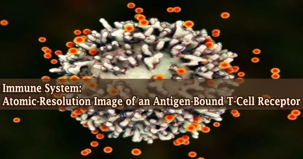
Our immune system’s specialized T cells are used to combat cancer cells and infectious illnesses. These unique white blood cells have a receptor that can identify antigens on their surface.
Biochemists and structural biologists from Goethe University Frankfurt, together with researchers from the University of Oxford and the Max Planck Institute of Biophysics, were able to see the entire T-cell receptor complex with bound antigen for the first time using cryo-electron microscopy.
Thus, they have contributed to our understanding of a fundamental mechanism that may open the door to creative therapeutic strategies that focus on serious diseases. Vertebrate immune systems are strong defenses against foreign infections and malignant cells.
T cells are crucial in this situation. They are equipped with a unique receptor on their surface known as the T-cell receptor, which is capable of recognizing antigens, which are tiny protein fragments from bacteria, viruses, and diseased or malignant bodily cells and are delivered by specialized immune complexes.
Thus, the T-cell receptor plays a key role in making the distinction between “self” and “foreign.” A signaling cascade inside the T cell is activated after an appropriate antigen binds to the receptor, “arming” the cell for the assigned duty.
The T-cell receptor is one of the most thoroughly studied receptor protein complexes, yet how this signaling pathway is activated has remained a mystery up until now.
Our structure is a blueprint for future studies on T-cell activation. In addition, it’s an important stimulus for employing the T-cell receptor in a therapeutic context for treating infections, cancer, and autoimmune diseases.
Robert Tampé
Many surface receptors change their spatial structure following ligand binding to transmit signals into the cell’s interior. The T-cell receptor was previously thought to be a part of this pathway.
Researchers led by Lukas Sušac, Christoph Thomas, and Robert Tampé from the Institute of Biochemistry at Goethe University Frankfurt, in collaboration with Simon Davis from the University of Oxford and Gerhard Hummer from the Max Planck Institute of Biophysics, have now succeeded for the first time in visualizing the structure of a membrane-bound T-cell receptor complex with bound antigen.
The initial hints about the activation mechanism come from a comparison between the structure of a receptor linked to antigen and one that is not.
The T-cell receptor utilized in immunotherapy for the treatment of melanoma was selected for the structural investigation because it has been specifically tailored to bind its antigen as firmly as possible.
Isolating the entire assembly of antigen receptors from the cell membrane, which consists of eleven distinct subunits, posed a unique difficulty on the path to structure determination.
“Until recently, nobody believed that it would be possible at all to extract such a large membrane protein complex in a stable form from the membrane,” says Tampé.
Once this was accomplished, the researchers employed a cunning technique to extract the functional receptors from the preparation that had made it through the procedure unscathed. By exploiting the potent interaction between the receptor complex and the antigen, they were able to “fish” one of the most crucial immune receptor complexes.
The subsequent images collected at the cryo-electron microscope delivered groundbreaking insights into how the T-cell receptor works, as Tampé summarises: “On the basis of our structural analysis, we were able to show how the T-cell receptor assembles and recognises antigens and hypothesise how signal transduction is triggered after antigen binding.”
Their findings show that the major surprise is that the spatial structure of the receptor does not appear to alter significantly after antigen binding because it was essentially unchanged with and without an antigen.
How antigen binding might alternatively result in T-cell activation is the last remaining mystery. After antigen binding, the co-receptor CD8 is known to move toward the T-cell receptor and induce the transfer of phosphate groups to its intracellular portion.
According to the researchers, this results in the creation of structures that block phosphate group-cleaving enzymes (phosphatases). In the absence of these phosphatases, the phosphate groups at the T-cell receptor stay stable and can initiate the subsequent stage of the signaling cascade.
“Our structure is a blueprint for future studies on T-cell activation,” Tampé is convinced. “In addition, it’s an important stimulus for employing the T-cell receptor in a therapeutic context for treating infections, cancer, and autoimmune diseases.”


