Researchers discovered significant changes in the brain structure of fetuses exposed to alcohol compared to healthy controls in the first MRI-based study of pre-natal alcohol exposure. The study’s findings were presented today at the annual meeting of the Radiological Society of North America (RSNA).
“Fetal alcohol syndrome is a global problem in countries where alcohol is widely available,” said Gregor Kasprian, M.D., associate professor of radiology at Austria’s Medical University of Vienna. “It is estimated that 9.8 percent of all pregnant women consume alcohol during their pregnancy, and this figure is likely to be underestimated.”
Fetal alcohol syndrome is the most severe form of a group of conditions known as fetal alcohol spectrum disorders, which are caused by prenatal alcohol exposure. Babies born with fetal alcohol spectrum disorders may have unique physical characteristics, learning disabilities, behavioral issues, or speech and language delays. According to Dr. Kasprian, one in every 70 pregnancies involving alcohol results in fetal alcohol syndrome.
For the study, we enlisted the help of 500 pregnant women who had been referred for a fetal MRI for medical reasons. “There have been many postnatal studies on infants who have been exposed to alcohol. We wanted to see how early changes in the fetal brain can be detected as a result of alcohol exposure.
Dr. Gregor Kasprian
“There have been many postnatal studies on infants who have been exposed to alcohol,” Dr. Kasprian explained. “We wanted to see how early changes in the fetal brain can be detected as a result of alcohol exposure.”
For the study, researchers enlisted the help of 500 pregnant women who had been referred for a fetal MRI for medical reasons. According to an anonymous questionnaire, 51 of the women admitted to drinking alcohol while pregnant. The Pregnancy Risk Assessment Monitoring System (PRAMS), a surveillance project of the Centers for Disease Control and Prevention and health departments, and the T-ACE Screening Tool, a measurement tool of four questions that identify risk drinking, were used as questionnaires.
“We provided a safe environment in which women could feel comfortable answering the questions honestly,” Dr. Kasprian explained.
After some fetal MRIs were discarded due to structural brain anomalies and/or poor image quality, the final study group included 26 fetal MRI exams from 24 alcohol-positive fetuses and a control group of 52 gender- and age-matched healthy fetuses. Fetuses ranged in age from 20 to 37 weeks at the time of imaging.
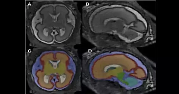
The researchers used super-resolution imaging to reconstruct each fetal brain from a single dataset. They then computed total brain volume and segment volumes of specific brain compartments after analyzing 12 different brain structures.
“One of the main strengths of our study is that we looked at so many smaller sub-compartments of the brain,” said co-author Marlene Stuempflen, M.D., a scientific researcher at the Medical University of Vienna.
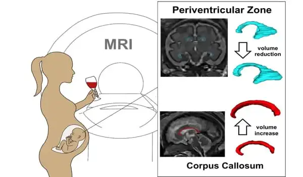
The statistical analysis revealed two significant differences in the alcohol-exposed fetuses compared to healthy controls: an increase in corpus collosum volume and a decrease in periventricular zone volume.
“This is the first time that a prenatal imaging study has been able to quantify these early alcohol-related changes,” said Dr. Stuempflen. The corpus collosum is the main connection between the two hemispheres of the brain. Dr. Stuempflen noted that this very central structure is affected because the clinical symptoms of fetal alcohol spectrum disorders are highly heterogeneous, or diverse, and cannot be pinpointed to a specific substructure of the brain.
“Changes found in the periventricular zone, where all neurons are born, reflect a global effect on brain development and function,” she explained.
The researchers were surprised to find a thicker corpus collosum in the alcohol-positive fetuses because the corpus collosum is thinner in infants with fetal alcohol spectrum disorders.
“It appears that alcohol exposure during pregnancy sets the brain on a path of development that deviates from a normal trajectory,” Dr. Kasprian explained. “Fetal MRI is a very powerful tool for characterizing brain development not only in genetic conditions, but also in acquired conditions caused by toxic agent exposure.”


