The research group at the Department of Robotics and Mechatronics Engineering at DGIST, under the direction of Professor Hongsoo Choi, has created a microrobot that can slice hippocampal tissues and form neural networks in an ex vivo setting.
The feasibility of using a microrobot to analyze structurally and functionally connected neural networks during cell delivery and transplantation in an in vitro environment has been confirmed through joint research with the team led by Dr. Jongcheol Rah from the Korea Brain Research Institute. The research, which was published in the journal Advanced Materials, is anticipated to have applications in a number of industries, including neural networks, cell therapy products, and regenerative medicine.
Various technologies involving microrobots capable of precise, minimally invasive cell delivery have been gaining popularity in recent years. Cell therapy products and cell delivery technology have been created to regenerate nerve cells damaged by diseases. Previous investigations using microrobots to deliver cells and connect neural networks only confirmed the structural and functional connections between cells at the cellular level.
“Using electrophysiological investigation, we demonstrated that a microrobot and mouse brain nerve tissues can be functionally coupled.”
Dr. Choi of DGIST
Microrobots, which can be used to practically apply neural network connections, were used in the research led by Professor Choi. Microrobots were used in this technology to analyze functionally connected neural networks in an ex vivo environment and deliver cells; the experiment was done on lab mice’s brain tissue.
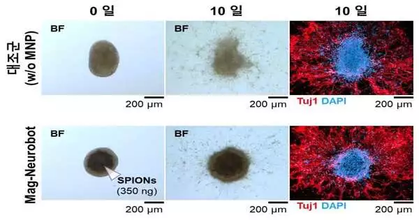
To create the Mag-Neurobot in a three-dimensional spherical form, the research team first fused superparamagnetic iron oxide nanoparticles to the main nerve cells of the lab mouse’s hippocampus. The robot’s exterior was covered in magnetic nanoparticles, allowing it to respond to magnetic fields from outside to move to a desired location. A biocompatibility test was also used to confirm the robot’s safety. In this test, nerve cell growth was not hampered by the robot’s magnetism.
The microrobot was inserted into the mouse’s hippocampus tissue section by the research team using magnetic field control. Through immunofluorescence staining, the team discovered that the cells in the microrobot and the cells in the tissue section of the hippocampus were structurally linked by neurites.
Additionally, to see if the nerve cells delivered by the microrobot exhibit typical electrophysiological traits, a microelectrode array (MEA) was used to stimulate the nerve cells within the microrobot. It was established that nerve cells within the tissue section of the hippocampus typically act as a conduit for electric signals.
The study group thus verified that the nerve cells delivered by the microrobot could actually function to form cells and neural networks inside the tissue section of a lab mouse’s hippocampus. The group also showed that the microrobot was capable of forming artificial neural networks and delivering nerve cells.
We have demonstrated through an electrophysiological analysis that a mouse brain’s nerve tissues and a microrobot can be functionally connected, according to Choi of DGIST.
“The technology developed in this study is anticipated to be used for verifying a precisely targeted treatment in the fields of cell therapy and neurological disorders.”.
More information: Eunhee Kim et al, A Neurospheroid‐Based Microrobot for Targeted Neural Connections in a Hippocampal Slice, Advanced Materials (2023). DOI: 10.1002/adma.202208747


