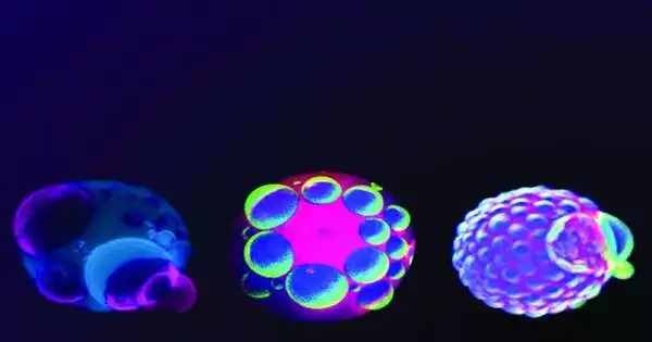
A living cell is packed with enormous, complex particles. Our understanding of how cells manage their essential biochemistry in a crowded space could be enhanced by new research on how these molecules could spontaneously organize themselves. This exploration may likewise reveal insight into how the primary living frameworks showed up and how they advanced their intricacies.
Eukaryotic cells contain coordinated designs, or organelles, limited by a lipid film. A model is the mitochondria, which produce energy in cells. As of late, researchers have found that notwithstanding these organelles, gatherings of particles can immediately frame into impermanent organelles, without a layer, to do some particular capability.
“There might be basic actual systems to make particular ‘creator organelles’ on request,” said Atul Parikh, teacher of biomedical design at the College of California, Davis.
“Coupling to the cell boundary prematurely stops phase separation and results in a droplet mosaic.” Intriguingly, these 3D droplets inside the vesicle reconfigure molecules in the 2D membrane, indicating to the exterior of the vesicle.”
Atul Parikh, professor of biomedical engineering at the University of California, Davis.
Utilizing an improved model of a cell, Parikh’s lab has found how combinations of polymers can parse into stage-isolated drops, similar to oils in an astro light, and that these beads communicate with the cell film surprisingly, including influencing the outside construction of the cell. The work is distributed July 6 in Nature Science.
Surface bubbling and liquid-liquid phase separation in polyethylene glycol (PEG) and dextran-containing vesicles. Credit: Wan-Chih Su, UC Davis
Wan-Chih Su, a graduate student at UC Davis who collaborated with Parikh, developed artificial vesicles that were approximately the same size as a living cell. These are basically rises with an engineered film containing water with two polymers broken down in it. The two polymers disintegrate in water, but they repel one another, so assuming they were combined as one and passed on to themselves, they would isolate into two stages, similar to an unmixed plate of mixed greens dressing.
As expected, Su and Parikh observed that when they removed water from the vesicles, the polymers began to form distinct droplets. However, rather than advancing to increasingly large drops, they observed that development came from connections between the polymer drops and within the vesicle layer, making a mosaic of beads.
Motioning beyond the cell
These collaborations likewise meaningfully affected the outside of the vesicle, causing a percolating or ‘blebbing’ impact. This appears to be like an impact found in living cells under certain conditions.
“Coupling to the cell limit rashly stops stage detachment and makes a mosaic of drops.” Interestingly, these three-dimensional droplets inside the vesicle reorganize molecules in the two-dimensional membrane, signaling to the outside of the vesicle,” Parikh stated. The scientists are sure that the peculiarity is, by and large, relevant and not intended for this specific mix of atoms.
The work shows how absolutely actual cooperations—hhow polymers repel or draw in one another—ccan lead to complex associations in an improved cell-like framework, Parikh said.
“We’re explaining the physical and synthetic standards behind science,” he said. “It could express something about how life might have occurred in any case.”
Parikh and associates intend to expand the work to include additional perplexing frameworks, including living cells.
Extra creators on the paper are Douglas Gettel, UC Davis; James Ho, C.S., Nanyang Mechanical College, Singapore; Christine Keating, and Andrew Rowland, both from Pennsylvania State University.
More information: Wan-Chih Su et al, Kinetic control of shape deformations and membrane phase separation inside giant vesicles, Nature Chemistry (2023). DOI: 10.1038/s41557-023-01267-1


