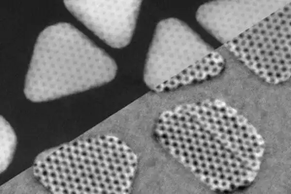
Researchers have mapped magnetic fields in three dimensions, which is a significant step toward resolving the “grand difficulty” of disclosing 3D magnetic configuration in magnetic materials. The findings have implications for increasing diagnostic imaging and storage device capacity.
The University of New Hampshire researchers have mapped magnetic fields in three dimensions, which is a significant step toward tackling what they call the “grand challenge” of disclosing 3D magnetic configuration in magnetic materials. The findings have implications for increasing diagnostic imaging and storage device capacity.
“The number three truly signifies a milestone in our discipline,” Jiadong Zang, associate professor of physics, stated. “Our brain exists in three dimensions. It’s amusing that all of our electronic devices are two-dimensional. They function poorly in comparison to our brains.”
The study, which was just published in the journal Nature Materials, presents the results of three years of high-performance numerical simulations mapping a three-dimensional structure of a 100 nm magnetic tetrahedron sample using only three electron beam projection angles. As an example, Zang cites computerized tomography medical imaging, or CT scans. Instead of using several X-ray beams to map tissues in the body, the same images might be created with only three beams.
The number three truly signifies a milestone in our discipline. Our brain exists in three dimensions. It’s amusing that all of our electronic devices are two-dimensional. They function poorly in comparison to our brains.
Jiadong Zang
One potential use for this collaborative research is reducing electron beam exposure in fast three-dimensional magnetic imaging. The discoveries of the researchers have implications for boosting the storage capacity of magnetic memory devices, which are currently depositing circuits onto two-dimensional panels that are approaching maximum density. This study’s technology will be valuable for detecting and characterizing three-dimensional magnetic circuits.
The theoretical analysis was carried out by Zang and Alexander Booth, a former UNH doctorate student. The physical experiments were carried out by researchers from Japan and the University of Wisconsin. The contributions of Zang and Booth to this research were aided by funds from the US Department of Energy (DOE), Office of Science, Basic Energy Sciences (BES), under award number DE-SC0020221.

This study also emphasizes areas of interest in magnetic resonance (MR) technology and applications, notably those involving dynamic magnetic resonance imaging (MRI). Among an MRI device’s hardware systems, the magnet, radio-frequency (RF), and gradient systems merit special R&D attention. One potentially promising area of interaction between solid-state physicists, materials scientists, and the biomedical imaging research community is the development of suitable high-temperature superconductors (HTSs) and their integration into MRI magnets.
This chapter discusses high-speed imaging pulse sequences, with a focus on functional imaging, as well as image reconstruction methods; both are attractive areas of research. Significant attention is being paid to two areas where MRI has a distinct advantage: blood flow imaging and measurement, and functional neuroimaging based on dynamic changes in magnetic susceptibility.
The University of New Hampshire inspires creativity and affects lives in our state, nation, and world. More than 16,000 students from all 50 states and 71 countries study with an award-winning faculty in top-ranked programs in business, engineering, law, health and human services, liberal arts, and the sciences throughout more than 200 programs of study. UNH, a Carnegie Classification R1 university, collaborates with NASA, NOAA, NSF, and NIH, and won $260 million in competitive external funding in FY21 to further explore and define the frontiers of land, sea, and space.


