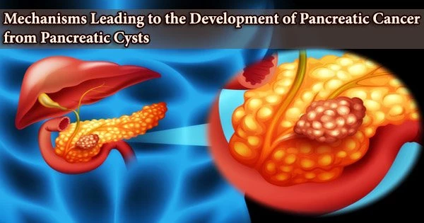
Because they are one of the few pancreatic cancer precursors that can be detected using radiologic imaging, pancreatic cysts have attracted a lot of attention recently.
A small percentage of these cysts, also known as pancreatic intraductal papillary mucinous neoplasms (IPMNs), will develop into invasive malignancy, despite the fact that the majority of them will remain benign. Although it has been hypothesized that the immune system contributes to the development of IPMNs into pancreatic cancer, the precise methods by which it does so remain unknown.
International researchers working under the direction of UC San Francisco have described the complete immunological environment and microbiome of pancreatic cysts as they develop from benign cysts to pancreatic cancer.
Their research, which was published on August 31 in Lancet Gastroenterology and Hepatology, may shed light on how neoplastic disease spreads and offer potential targets for immunotherapy.
“This will have far reaching implications on how we think about utilizing immunotherapies to treat certain types of pancreatic cancer, and also potentially inhibit the formation and progression of cancer from pancreatic cysts,” said senior author Ajay V. Maker, MD, FACS, FSSO, a pancreatic surgeon, chief of UCSF’s division of surgical oncology, and Maurice Galante Distinguished professor of surgical oncology.
The IPMNs’ immunological tumor microenvironment changes as the malignancy progresses. The cellular environment that surrounds a tumor and contains immune cells as well as a stroma that sustains other cells and tissues is known as the tumor microenvironment.
This will have far reaching implications on how we think about utilizing immunotherapies to treat certain types of pancreatic cancer, and also potentially inhibit the formation and progression of cancer from pancreatic cysts.
Ajay V. Maker
The microenvironment constantly interacts with a tumor, impacting the development of both healthy and malignant cells (dysplasia).
A cytotoxic immune response with a high concentration of CD8+T cells evolves into an immunosuppressive environment with a discernible inflammatory response as neoplasms advance from low-grade dysplasia to high-grade dysplasia and finally to invasive carcinoma.
The researchers hypothesize that therapies that promote cytotoxic T cells may be ideal for IPMNs with low-risk disease, while therapies that target regulatory T cells, myeloid-derived suppressor cells, and inhibitory macrophages may be helpful in slowing the progression of malignancy and treating high-risk disease.
Treating the immune tumor microenvironment in addition to IPMNs with an accompanying invasive carcinoma may stop the progression of IPMNs with a high risk of malignant transformation.
The researchers explain that the tumor microenvironment exhibits an increase in the levels of pro-inflammatory and cyst fluid cytokines with increasing quantities of dysplasia, which is suggestive of a T-cell immunological response.
According to the team’s findings, assessing and characterizing the immune response to IPMNs may enable early detection and maybe improve therapies for invasive disease prevention or treatment.
Further research on the tumor immune microenvironment of pre-invasive lesions is essential, according to the researchers, who also recommend comparing main-duct disease to branch-duct disease.
Funding:
Ajay V Maker’s research in tumor immunology is funded through the National Institutes of Health Method to Extend Research in Time award (R37CA238435), and he has the following pending or issued patents: WO2018183603A1 (PCT/US2018/025027) and US9757457B2.


