A joint examination group from DGIST drove by Teachers Jin Ho Chang and Jae Youn Hwang of the Branch of Electrical Designing and Software engineering has fostered the world’s most memorable laser checking microscopy innovation that empowers further and more itemized perception of organic tissues utilizing gas bubbles that are briefly created by ultrasound.
Optical imaging and helpful advances are broadly used in life science research and clinical practice. Nonetheless, because of the event of optical dispersing in the tissue, the light transmission is low. Thus, there exists an inborn limit in the picture securing and treatment of profound tissue. This altogether ruins the extension of the application field.
To defeat this, in 2017, Teacher Jin Ho Chang’s group imagined that it would be feasible to utilize micrometer-sized gas bubbles that are regularly seen when tissues are presented to extreme focus ultrasound. They fostered an innovation in view of the way that gas bubbles briefly made by ultrasonic waves cause optical dispersing in a similar course as the spread of episode light, hence expanding the entrance profundity of light.
“We were able to overcome the inherent constraints of existing optical imaging and therapy technologies through close collaboration with ultrasound and optical imaging experts.”
Professor Jin Ho Chang of the Department of Electrical Engineering and Computer Science at DGIST
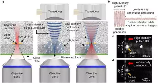
Besides, the joint examination group of Teachers Jin Ho Chang and Jae Youn Hwang zeroed in on growing the use of optical imaging innovation utilizing ultrasound-prompted gas bubbles. A confocal fluorescence magnifying lens is a gadget that specifically identifies fluorescence signals created at the central plane of light and gives high-goal, high-contrast pictures of microstructures like disease cells. It is the most broadly involved gadget in life science research attributable to its elite exhibition. Nonetheless, the shine of the light is obscured at profundities surpassing 100 µm inferable from the dispersing of light that happens inside the tissue, which altogether restricts the application and adequacy of confocal fluorescence microscopy.
To build the greatest imaging profundity of optical imaging modalities, for example, confocal fluorescence microscopy, photons comprising the illuminated light should not have a peculiarity where their spread course is twisted by light dispersing in tissues. Nonetheless, the recently evolved strategy in view of gas bubbles meagerly made by ultrasound was not an answer.
Hence, this joint examination group created ultrasound innovation to make an air pocket layer in the ideal region with thick gas rises (with a thickness of 90% or more) inside living tissue and to keep up with the produced gas rises while gaining the picture. In this gas bubble layer, the bending in the spread course of photons doesn’t happen.
Hence, it has been tentatively demonstrated that light centering is conceivable even in more profound natural tissues. Moreover, by applying this innovation (i.e., ultrasound-prompted tissue straightforwardness) to a confocal fluorescence magnifying lens, the UltraSound-incited Optical Clearing Microscopy (US-OCM), named in this review, was created without precedent for the world with an imaging profundity quite a bit longer than that of the regular confocal microscopy.
Specifically, the US-OCM created in this study made no harm the tissue since when the ultrasonic light was halted, the produced gas bubbles vanished, and the optical properties got back to before gas bubble age, proposing that it is innocuous to the living body.
Teacher Jin Ho Chang of the Branch of Electrical Designing and Software engineering at DGIST says that “through close cooperation with ultrasound and optical imaging specialists, we had the option to beat the innate limits of existing optical imaging and treatment advances.”
“The innovation got through this study will be applied to different optical imaging methods including multiphoton microscopy and photoacoustic microscopy notwithstanding a few optical treatments including photothermal treatment and photodynamic treatment. This would upgrade the use of existing advances by expanding their picture and treatment profundity.”
The consequences of this examination were distributed in Nature Photonics.
More information: Haemin Kim et al, Deep laser microscopy using optical clearing by ultrasound-induced gas bubbles, Nature Photonics (2022). DOI: 10.1038/s41566-022-01068-x
Journal information: Nature Photonics


