Fluorescence imaging can be led with long Stokes shift colors that limit crosstalk between the excitation source and fluorescent outflow to work on the sign to-foundation proportion. In any case, analysts actually structure basic, little atom colors with huge Stokes shift and close to infrared outflows. In another report currently distributed in Science Advances, Hao Chen and a group of researchers fostered a progression of styrene oxazolone colors (SODs) utilizing basic engineered techniques roused by the chromophore compound design of fluorescent proteins.
The colors displayed close infrared outflows with long Stokes shifts and little atomic weight. The most encouraging colors likewise showed fast renal discharge and blood-mind boundary passing properties. The bioengineers altered the mixtures for growth imaging, fluorescence picture directing a medical procedure, neurosurgery, and neurotic examination. The discoveries contribute a fundamental little atomic color classification to the old-style colors.
Stokes’ colors change as he grows older.
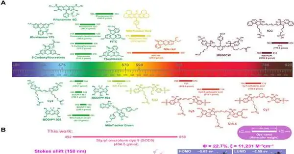
Fluorescence imaging is broad in preclinical biomedical examination, as well as clinical pathology and fluorescence imaging directed at a medical procedure. The simple, minimal expense stage offers minor light harm to the natural example for high location awareness. The biomedical use of fluorescent imaging relies upon the colors with basic elements, including retention and outflow profiles, ingestion coefficient, quantum yield, Stokes shift, and photochemical strength.
By and by, a couple of colors have shown ideal properties across all classes. The weighty crosstalk between the excitation and outflow light can bring about a somewhat low sign of to-foundation proportion. Thus, natural chemists plan to grow long Stokes shifts close to infrared colors for high sign to foundation proportion. In this work, Chen and the group depicted the main high-proficiency styrene oxazolone colors (SODs) as lengthy stirred up shift colors, to give another system for in vivo fluorescence imaging.
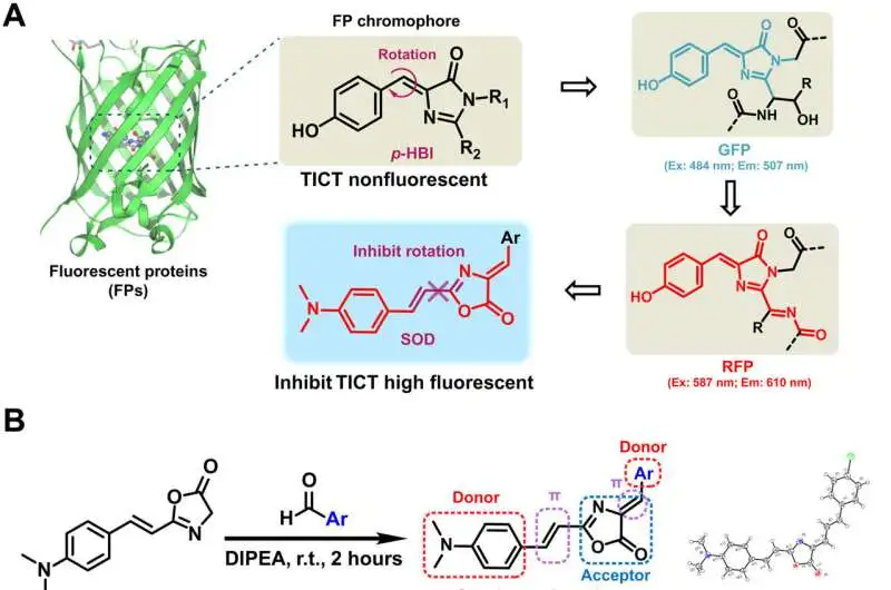
plan and union of SODs. (A) The chromophore compound designs of GFP and RFP (B) synthesis of SODs (left) and the gem design of SOD10 (right). DIPEA, N,N-Diisopropylethylamine. (C) The SOD color substance designsExcitation; Emulation; TICT, bent intramolecular charge move; r.t., room temperatureScience Advances (2022) is credited.DOI: 10.1126/sciadv.abo3289
Tests: Design, union, and portrayal of styrene oxazolone colors (SODs)
Fluorescence proteins are broadly concentrated in natural examination, where they share a similar center skeleton of an imidazolinone moiety and an exocyclic twofold cling to switch between the dim and splendid states. The scientists planned and blended a progression of previously unreported colors with styrene oxazolone as a key skeleton through a basic method at room temperature for good yields in two hours or less. They described the compound design of SODs utilizing standard 1H atomic attractive reverberation (NMR) and high-goal mass spectrometry spectra. The group recognized the spectroscopic properties of the colors in fluid media, where they noted solid fluorescence with great photostability because of the presence of an exocyclic twofold bond. The analysts determined the distinction in spectroscopic properties from the electrical properties and position of the substituents.
They hence showed what various elements meant for the optical properties of the SODs (styrene oxazolone colors) and analyzed the cytotoxicity of the colors from there on. They followed these tests with in vivo pharmacokinetics through fluorescence imaging, as well as in vivo uses of color for biomolecule naming. The outcomes showed how a few tests focusing on neurotic circumstances gathered more in growth cells than in typical cells, to feature their cancer-specific focusing properties.
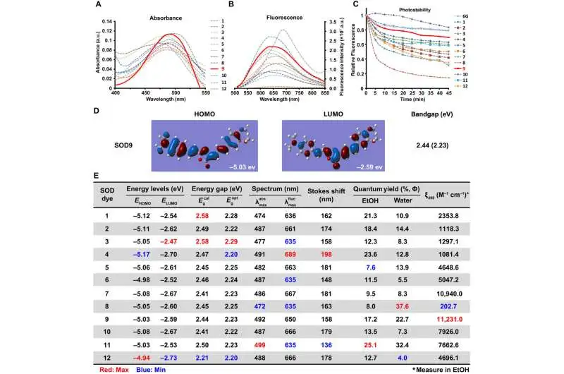
optical properties of SODs. The absorbance (A), fluorescence range (B), and photostabilities (C) of SODs were estimated in water with the grouping of 20, 12, and 10 M, separately (6G addresses rhodamine 6G). SOD9 advanced atomic orbital plots (HOMO and LUMO) based on the density useful hypothesis (DFT).(E) A list of the optical properties of SOD colorsRed is the greatest value, and blue is the base value of a similar section. EtOH, ethanol. Science Advances (2022) is credited.DOI: 10.1126/sciadv.abo3289
Biocompatibility of the intravenous infusion of the colored biomolecules
The scientists concentrated on the biocompatibility of the color after intravenous infusion to a mouse model and explored its impact on the inner organs through histology. The results showed the possibility of utilizing the new fluorescence test to recognize cancers and significant organ fluorescence imaging. The work stressed the best time for fluorescence picture-directed growth medical procedure to be an hour post-infusion and showed how the quick mind amassing of the color momentarily made it reasonable for cranial nerve dynamic imaging. The results featured the effect of the color atom for mind-neuron dynamic checking.
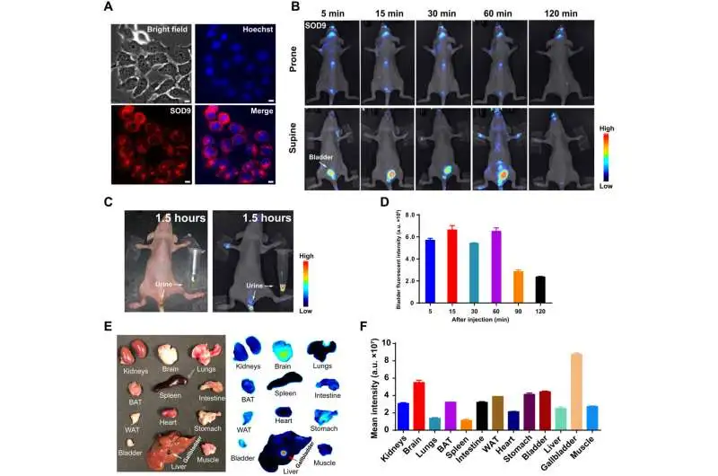
SOD9’s in vivo pharmacokinetics by fluorescence imaging. (A) SOD9 fluorescence imaging in NIH-3T3 cells (red) and convergence with the atomic stain Hoechst (blue). Scale bars, 10 μm. (B) Whole-body NIR imaging of bare mice (n = 3, inclined and recumbent situations) after intravenous infusion of SOD9 (2.5 mg/kg, 6.18 mol/kg). The sign was gathered in the 650-to 800-nm channel with an excitation of 500 nm. (C) Colored imaging (top) and fluorescence imaging (bottom) of bare mice with pee discharge 90 minutes after intravenous SOD9 infusion.(D) Comparison of bladder fluorescent forces at various time points after intravenous infusion of SOD9. Mistake bars imply SD (n = 3). (E) Ex vivo imaging of the significant organs analyzed subsequent to euthanizing creatures at 2 hours after an intravenous infusion of SOD9 (10 mg/kg). Left: a hued picture; right: fluorescence imaging. (F) Comparison of mean powers for the significant organs at 2 hours after intravenous infusion of SOD9. SD (n = 3) implies blunder bars.Credit: ScienceAdvances (2022). DOI: 10.1126/sciadv.abo3289
Viewpoint
Along these lines, Hao Chen and partners planned and fostered a progression of oxazolone analogs and determined their enhanced math. The subsequent color analogs showed a lower bandgap to add to a bigger Stokes shift, approximately 150 to 200 nm more prominent than customary fluorescence colors. The substituents and steric block impacts played a key part in impacting the spectroscopic properties of the colors. The results showed great optical and pharmacokinetic properties with high sign to-foundation proportion, fast leeway, and low harmfulness. The particles stunningly crossed the blood-mind boundary after intravenous infusion into mice to give areas of strength for a sign to picture neurons through confocal fluorescence imaging in vivo.
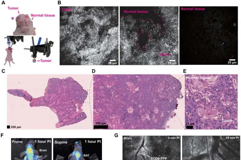
SOD9-TPP for fluorescence imaging for medical procedures, brain neuroimaging, and on-site pathologic examination. (A) Top: The hued picture of the orthotopic HNSCC mouse (SCC090; cancer set apart with the red pentagram). The arrangement’s variety of photographs of the confocal fluorescent endomicroscopy imaging-directed a medical procedure. Base right: The arrangement’s hued photograph of the confocal fluorescent endomicroscopy imaging of the resected tissue. (B) Confocal fluorescent endomicroscopy imaging of the analyzed HNSCC growth during fluorescence picture directed a medical procedure on mice 2 hours after SOD9-TPP (5.0 mg/kg, 6.13 mol/kg) intravenous infusion. Right, cancer; center, growth and typical tissue; left, ordinary tissue. H&E staining of HNSCC growth tissue areas (C). (D) A zoomed-in version of (C). (E) A zoomed-in version of (D). (F) Whole-body NIR imaging of bare mice (n = 3, inclined and recumbent situations) after intravenous infusion of SOD9-TPP (5.0 mg/kg, 6.13 mol/kg); SOD9-TPP was tracked down in the mind, BAT, and liver. (G) Different time focuses in vivo confocal fluorescent endomicroscopy imaging of mind neurons with the skull opened. 25 m scale bars (H) Imaging of significant organs using in vivo confocal fluorescent endomicroscopy with the midsection and chest opened. 25 m scale bars Credit: ScienceAdvances (2022). DOI: 10.1126/sciadv.abo3289
The group additionally changed the convention to permit staining of mitochondria in living cells with applications across growth imaging, fluorescence route, a medical procedure, and confocal endoscopy to recognize careful limits and forestall careful injury. The new procedures worked with the examination of cell morphology continuously, which improved the course of regular histological assessment with the extra ability to supplant customary techniques for staining like Hematoxylin and Eosin too. The colors are a formerly unreported compound that can be utilized for biomedical applications during fluorescence-directed medical procedures, with promising properties including high quantum proficiency, low cytotoxicity, fast discharge, and fluorescence imaging.
More information: Hao Chen et al, Bioinspired large Stokes shift small molecular dyes for biomedical fluorescence imaging, Science Advances (2022). DOI: 10.1126/sciadv.abo3289
Guosong Hong et al, Near-infrared fluorophores for biomedical imaging, Nature Biomedical Engineering (2017). DOI: 10.1038/s41551-016-0010
Journal information: Nature Biomedical Engineering


