According to current models of the visual system developed by neuroscientists, it encodes the positions of various objects similarly to how a camera would, representing the visual world in a similar way. But since the environment in which an animal lives is constantly changing, this change may also affect how visual information is processed.
This theory is supported by evidence recently gathered by scientists at the Institute of Science and Technology in Austria and the LMU in Germany, which demonstrates that the organization of neurons in the mouse retina is impacted by panoramic (i.e., wide-view) visual statistics such as variations in light intensity. Their discoveries, which were reported in Nature Neuroscience, may significantly advance current knowledge of the visual system and its development.
According to Maximillian Jösch, one of the study’s researchers, “every living organism must adapt to its environment in order to survive.”. “Such adaptations should also take place in the brain’s computations, for example, to extract relevant information and ignore unimportant data. We set out to test this hypothesis by utilizing the most notable visual changes that have been systematically observed in nature: the gradient of light intensity and contrast levels from the ground to the sky. We wanted to know if the mouse visual system had evolved to take these constraints into account.
“We can visualize that activity by implanting a fluorescent indicator in each neuron.” When calcium enters, the fluorescence changes. These fluctuations in fluorescence may be captured using a sensitive camera, allowing us to deduce how the neuron responds to diverse visual stimuli over the entire retina.”
Maximillian Jösch, one of the researchers who carried out the study,
Jösch and his colleagues created a brand-new optical imaging method to study how sensory space is organized in the mouse retina’s receptive fields in relation to the scenes that mice are viewing. They can measure and monitor the activity of thousands of neurons in a single retina at once using this method.
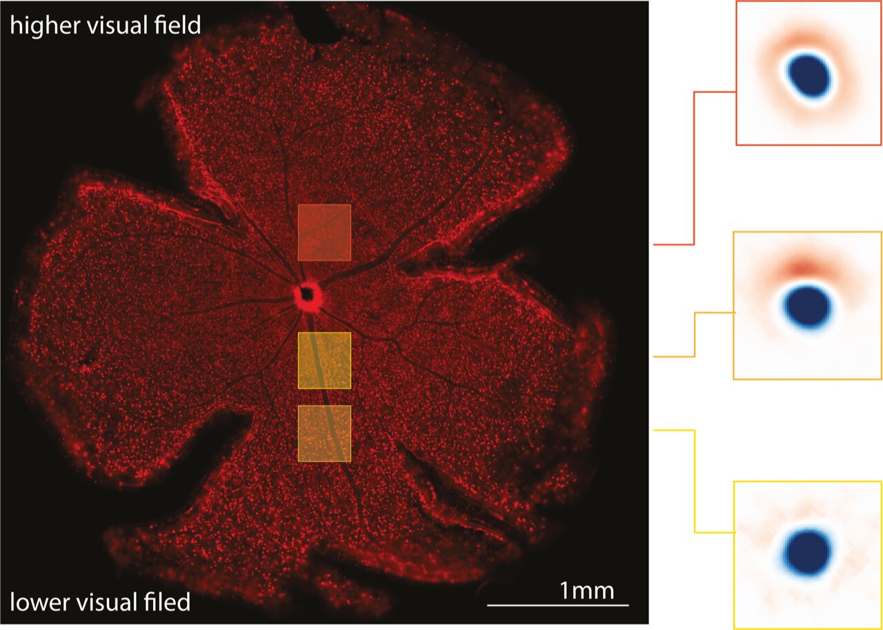
Left: A mouse retina with retinal neurons stained in red. Right: changes in the visual properties of the retinal cells across elevation. Credit: Divyansh Gupta
“How does our optical technique work? When a retinal neuron is active, sending electrical pulses to the brain, ions flow inside the cell, e.g., calcium,” Jösch cited. The incorporation of a fluorescent indicator into each neuron allows us to see that activity. Fluorescence changes as calcium enters. Using a sensitive camera to record these fluorescence changes, we can determine how the neuron reacts to various visual stimuli throughout the entire retina.”.
On mouse retinas that had been removed, the researchers ran their experiments. The mouse retina, like those of the majority of mammals, lacks the fovea, a tiny depression in the retina that enables humans and other primates to see in high definition. The fovea, which accounts for less than 1% of the entire human retina, is known to play a significant role in the visual perceptions of which humans are more conscious. Visual perceptions are also influenced by the remaining 99 percent of the human retina, many of which seem to be unconscious processes. So, from a human-centric standpoint, this study focuses on the processing that occurs in the latter 99 percent.
Jösch and his coworkers discovered that the calculations made by neurons in the mouse retina varied based on the panoramic visual statistics of what that region of the retina typically sees in daylight. This corroborates their original claim that the visual system is not inherently homogenous but rather adapted to the outside world.
Surprisingly, Jösch said, “we discovered that retinal neurons are more likely to alert the rest of the brain when a stimulus change is unexpected. Importantly, the unexpected is dependent on whether the neuron is looking up at the sky or down at the ground. As a result, retinal circuits changed their characteristics gradually from the lower to the higher visual field to more effectively represent the outside world.”.
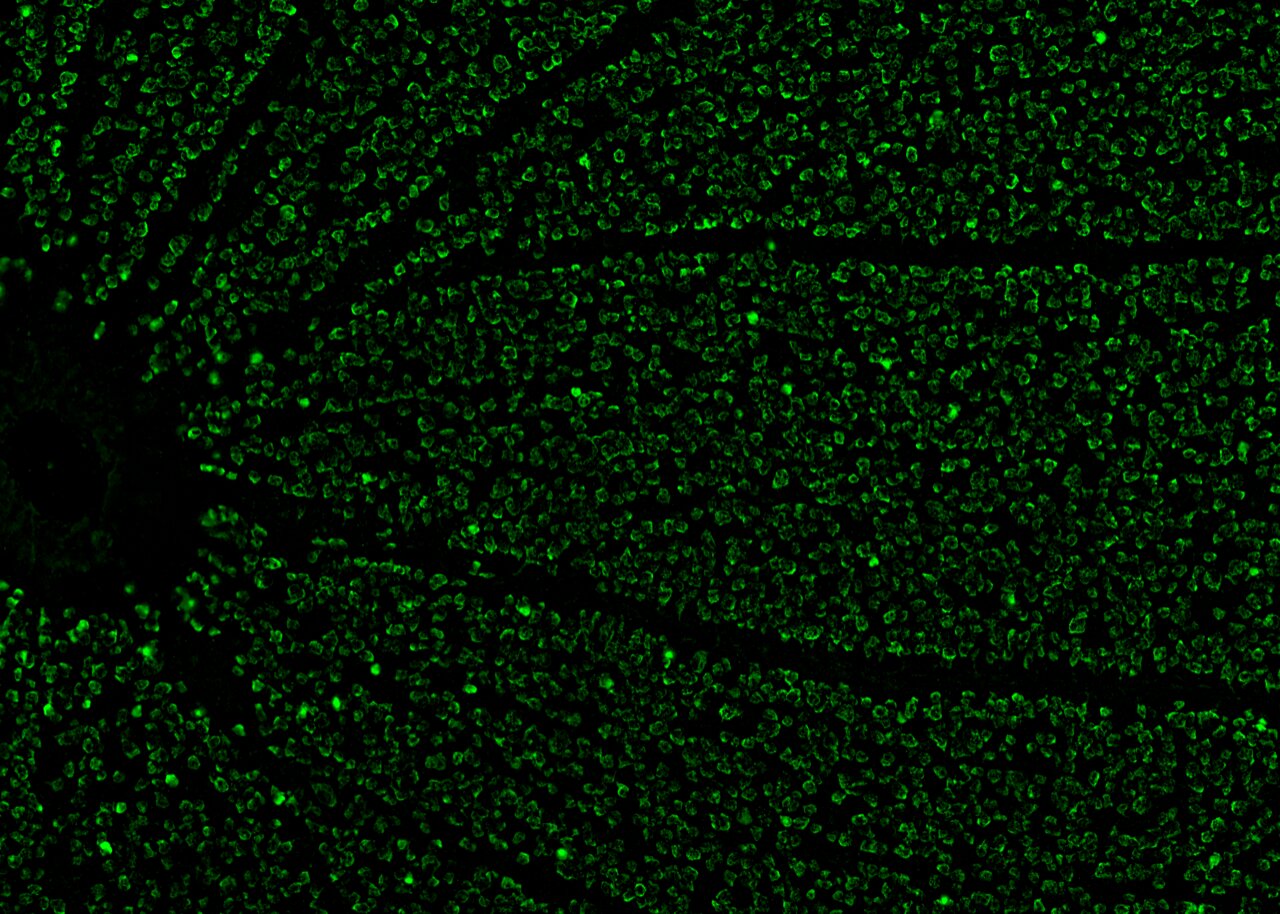
A mouse retina and the retinal neurons in green. Credit: Divyansh Gupta
Overall, the research team’s findings point to the possibility that the panoramic nature of natural scenes influences how various processing methods are organized in various retinal regions. This enhances earlier theories of the visual system by showcasing its adaptable and dynamic nature.
We frequently assume that the visual system is homogeneous, or, to put it another way, that the visual world is viewed through a camera that measures each position uniformly. But the natural world around us is not the same; it gradually shifts from the ground to the sky. As a result, a system that has evolved to survive in nature should take this into account. Our findings suggest that the visual system of living things has evolved to deal with environmental restrictions in order to increase the effectiveness of their neuronal code.”.
The recent work by Jösch and his colleagues may encourage other groups to investigate how panoramic statistics or other visual components affect cell organization in the retina in the future, improving our understanding of vision in general.
Jösch continued, “We are now investigating how similar adaptations change when the context changes, for example, when adapting to different light levels occurring during the day or at night.
More information: Divyansh Gupta et al, Panoramic visual statistics shape retina-wide organization of receptive fields, Nature Neuroscience (2023). DOI: 10.1038/s41593-023-01280-0


