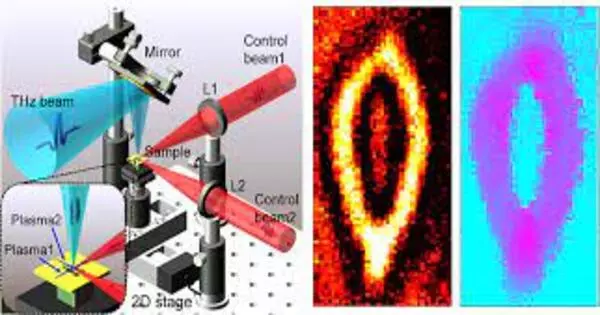
As a novel far-infrared review strategy, the advancement of terahertz (THz) imaging innovation has drawn significant consideration lately. With the remarkable properties of THz radiation, for example, non-ionizing photon energies and wide phantom data, this imaging procedure has shown strong application potential in numerous major exploration and modern fields. Nonetheless, the goal of THz imaging is generally restricted because of its long frequency. The presentation of optical close-field procedures can extraordinarily upgrade the goal, but it is generally fundamental to expect that a THz source or indicator approach the example from quite a distance. For delicate or fluid materials in biomedical detecting and compound examination, these examples might be effortlessly harmed and the THz source or locator might be tainted in conventional THz close field strategies. Consequently, it is actually a test to accomplish THz close-field microscopy in more extensive application fields.
In another paper distributed in Light: Science and Applications, a group of researchers, led by Professors Xin-ke Wang and Yan Zhang from Beijing Key Laboratory of Metamaterials and Devices, Key Laboratory of Terahertz Optoelectronics Ministry of Education, Department of Physics, Capital Normal University, Beijing, China, and colleagues, have fostered another THz close field microscopy to accomplish THz sub-frequency imaging without moving toward the example with any gadgets.
In this THz close field procedure, a cross-fiber was framed by two cross air-plasmas, which opened a powerful gap to balance the force of a THz shaft on an example surface. Whenever the cross-fiber was adequately close to the example surface, THz imaging with the goal of many microns was satisfied. Taking advantage of the benefits of this method, the impediment of the example decision was actually eliminated in conventional THz close field imaging and test harm from the cross-fiber was limited.
To check the exhibition of the strategy, four different types of materials were estimated and their THz sub-frequency pictures were effectively procured, including a metallic goal test outline, a semiconductor chip, a plastic example, and an oily spot. What’s more, the method is likewise reasonable on a basic level for an epitomized test, assuming that its bundling is straightforward to THz and noticeable light. Consequently, it may very well be guessed that the revealed technique will essentially widen the uses of THz close field microscopy, e.g., biomedical detection and compound review.
Conversation
Compared with past reports on THz close field microscopy5,6,13,14,38,39, the benefit of our strategy was not the goal ability, but rather the way that no THz identifier or source was near the example surface, guaranteeing that the method was relevant to various sorts of tests, like metals, semiconductors, colloid, and fluidic materials. In the past reports, it was fundamental that a THz finder or source moved toward the example with a metallic aperture40, dissipating tip41, film photomodulator38, spintronic THz emitter14, electro-optic crystal39, or miniature organized photoconductive antenna9. Delicate materials could hence be easily harmed and the THz locator or source could be tainted. Here, we simply had to change the level h and the beat energies of the control shafts to forestall test harm, as exhibited in the oily spot imaging. The plan is also appropriate on a fundamental level for a standard test if its bundling is straightforward to THz and visible light. Besides, transmission and reflection estimation modes could be upheld at the same time, which extends the testing ability. In conclusion, it is reasonable to expect that our technique will significantly broaden the applications of THz close-field microscopy.
There are multiple ways of working on the strategy. In the future, high level diffraction optical elements42,43, and metasurface devices44,45 could be used to tweak the wave front of a control shaft, and a miniature plasma in three dimensions could be formed by just a control bar. Along these lines, the goal could be further advanced by diminishing the size of the plasma. The SNR of the ongoing framework was as yet confined, which impacted the imaging quality. High level THz producers, such as natural crystals46, lithium niobate47, and spintronic emitters48, could be used in ongoing upgrades. Besides, high-level computerized picture handling calculations could be used to all the more proficiently remove THz close-field data.
Taking everything into account, another THz close-field microscopy method was illustrated. It depended on an air-plasma dynamic opening that didn’t need a close way to deal with an example by a THz finder or source to acquire a semi-sub-frequency goal (roughly /2). Close field imaging of metallic, semiconductor, plastic, and oily examples was illustrated. The goal qualities were explored exhaustively through the blade edge strategy, and an opening transmission model was utilized to analyze the actual instrument. In general, the strategy opens another heading for THz microscopy for central exploration and industry.


