There is growing evidence to suggest that disrupted flow of brain fluid, or cerebrospinal fluid (CSF), may underlie certain neurodevelopmental disorders. CSF is a clear, colorless liquid that surrounds the brain and spinal cord, providing nutrients, cushioning, and waste removal. It is constantly produced and reabsorbed by the brain, and its flow is regulated by complex mechanisms.
Researchers discovered that fluid that circulates through the brain flows to areas critical for normal brain development and function in rodents, implying that disruptions in its circulation may play an unnoticed role in neurodevelopmental disorders.
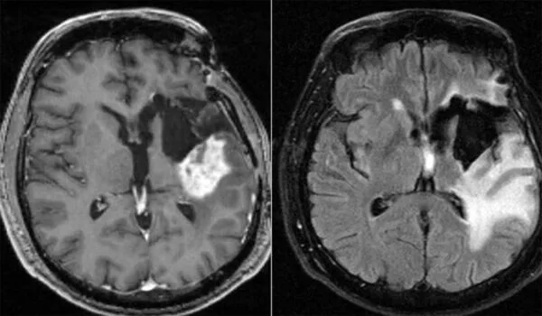
The brain floats in a sea of fluid that protects it from injury, supplies it with nutrients, and removes waste. Disruptions in the normal ebb and flow of the fluid have been linked to neurological conditions such as Alzheimer’s disease and hydrocephalus, a disorder characterized by an accumulation of fluid around the brain.
Researchers from Washington University School of Medicine in St. Louis developed a new technique for tracking fluid circulation patterns in the brain and discovered that it flows to areas critical for normal brain development and function in rodents. The researchers also discovered that circulation appears abnormal in young rats with hydrocephalus, a condition linked to cognitive deficits in children.
The findings, published in Nature Communications, suggest that the fluid that surrounds the brain, known as cerebrospinal fluid, may play an unnoticed role in normal brain development and neurodevelopmental disorders.
Disordered cerebrospinal fluid dynamics could be responsible for the changes in brain development we see in children with hydrocephalus and other developmental brain disorders.
Jennifer Strahle
“Disordered cerebrospinal fluid dynamics could be responsible for the changes in brain development we see in children with hydrocephalus and other developmental brain disorders,” said senior author Jennifer Strahle, MD, an associate professor of neurosurgery, of pediatrics, and of orthopedic surgery. As a pediatric neurosurgeon, Strahle treats children with hydrocephalus at St. Louis Children’s Hospital.
“A variety of neurologic disorders, including hydrocephalus, are associated with developmental delays in young children. We don’t know what’s causing the developmental delays in many of these conditions. It is possible that in some of these cases, the brain regions through which cerebrospinal fluid circulates have altered function.”
A great deal of research has been done to map the drainage of cerebrospinal fluid in adult brains. However, how cerebrospinal fluid interacts with the brain is unknown. Because young children do not yet have the mature drainage pathways of adults, cerebrospinal fluid pathways in the brain are likely to vary with age.
Strahle; first author Shelei Pan, an undergraduate student; and colleagues developed an X-ray imaging technique using gold nanoparticles that allowed them to visualize brain circulation patterns in microscopic detail. Using this method on young mice and rats, they showed that cerebrospinal fluid enters the brain through small channels primarily at the base of the brain, a route that has not been seen in adults. In addition, they found that cerebrospinal fluid flows to specific functional areas of the brain.
“These functional areas contain specific collections of cells, many of which are neurons, and they are associated with major anatomic structures in the brain that are still developing,” Strahle said. “Our next steps are to understand why cerebrospinal fluid is flowing to these neurons specifically and what molecules are being carried in the cerebrospinal fluid to those areas. There are growth factors within the cerebrospinal fluid that may be interacting with these specific neuronal populations to mediate development, and the interruption of those interactions could result in different disease pathways.”
Further research revealed that hydrocephalus reduces cerebrospinal fluid flow to specific neuron clusters. Strahle and colleagues looked into a type of hydrocephalus that affects premature babies. Premature babies are at risk of brain bleeding around the time of birth, which can result in hydrocephalus and developmental delays. Strahle and colleagues induced in young rats a process that resembled that of premature babies. The tiny channels that carry cerebrospinal fluid from the brain’s outer surface into the middle were fewer and shorter after three days, and circulation to 15 of the 24 neuron clusters was significantly reduced.
“There hasn’t been much research into the idea that cerebrospinal fluid can regulate neuronal function and brain development,” Strahle said. “It is common to see cognitive dysfunction in the setting of hydrocephalus, even after the excess fluid has been successfully drained. The disordered cerebrospinal fluid dynamics to these functional brain regions may eventually affect brain development, and normalizing flow to these areas is a potential approach to reducing developmental problems. It’s an exciting field, and we’re only scratching the surface of what cerebrospinal fluid can do.”
While the exact mechanisms underlying the relationship between disrupted CSF flow and neurodevelopmental disorders are still being investigated, the findings suggest that CSF flow may be an important factor to consider in understanding the pathophysiology of these conditions. This research may also lead to the development of new diagnostic and treatment approaches that target CSF flow and associated mechanisms.