Uncommon perspectives on the inside of cells and other nanoscale structures are currently possible on account of advancements in extension microscopy. The advancements could help with future knowledge in neuroscience, pathology, and a variety of other organic and clinical fields.
In the paper “Amplify is a general sub-atomic securing procedure for extension microscopy,” distributed Jan. 2 in the journal Nature Biotechnology, associates from Carnegie Mellon College, the College of Pittsburgh, and Brown College portray new conventions for Amplify.
“Amplify can be a powerful and open tool for the biotechnology community,” said Yongxin (Leon) Zhao, the Eberly Family Vocation Improvement Academic Partner of Natural Sciences.
Zhao’s Biophotonics Lab is a forerunner in the field of empowering super-goal imaging of organic examples through genuinely growing examples in a cycle known as development microscopy. Through the cycle, tests are implanted in a swellable hydrogel that homogenously grows to expand the distance between particles, permitting them to be seen in a more noteworthy manner. This enables nanoscale natural designs that could previously only be seen with expensive high-resolution imaging methods to be seen with standard microscopy instruments.
“We overcome certain long-standing expansion microscopy problems. Magnify’s uniform technique for keeping the tissue’s biomolecules, such as proteins, nucleus fragments, and carbohydrates, within the larger sample is one of its primary selling advantages.”
Yongxin (Leon) Zhao, the Eberly Family Career Development Associate Professor of Biological Sciences.
Amplify is a variation of development microscopy that permits specialists to utilize another hydrogel equation, imagined by Zhao’s group, that holds a range of biomolecules, offers a more extensive application to different tissues, and builds the extension rate up to multiple times directly or 1,300 folds of the first volume.
A video shows kidney cells. Developmental microscopy (ExM) provides unique views of cell interiors. The emerging super-objective imaging procedure is based on physical, rather than optical, amplification.CMU’s Zhao Biophotonics Lab advances the development rate and allows for the visualization of various tissues in 3D.
“We defeated a portion of the longstanding difficulties of development microscopy,” Zhao said. “One of the primary selling points for Amplify is the widespread methodology to keep the tissue’s biomolecules, including proteins, core bits, and sugars, inside the extended example.”
Zhao said that safeguarding different organic parts matters in light of the fact that past conventions required disposing of numerous different biomolecules that kept tissues intact. Yet these particles could contain significant data for analysts.
“Previously, to make cells truly expandable, you really wanted to utilize compounds to process proteins, so eventually, you had a vacant gel with names that showed the area of the protein of interest,” he said. With the new strategy, the particles are held together, and different kinds of biomolecules can be marked in a solitary example.
“Previously, it resembled having single-decision questions. To mark proteins, that would be the variant one convention. “To name cores, then that would be an alternate form,” Zhao said. “If you had any desire to do synchronous imaging, it was troublesome.” Presently, with Amplify, you can pick numerous things to mark, like proteins, lipids, and carbs, and picture them together.
Lab scientists Aleksandra Klimas, a postdoctoral specialist, and Brendan Gallagher, a doctoral understudy, were the first co-authors on the paper.
“This is an available method for imaging examples in high-speed imaging,” Klimas said. “Generally, you want costly hardware and explicit reagents and preparation.” In any case, this technique is comprehensively relevant to many sorts of test arrangements and can be seen with standard magnifying lenses that you would have in a science research center.
Gallagher, who has experience with neuroscience, said their objective was to make the conventions as viable as possible for analysts who could profit from embracing the Amplify as a feature of their tool compartments.
“One of the key ideas that we attempted to remember was to meet scientists where they are and have them change as few things in their conventions as could be expected,” Gallagher said. “It works with various tissue types, obsession strategies, and even tissue that has been safeguarded and put away.” It is entirely adaptable, in that you won’t be guaranteed to have to upgrade; it explores different avenues regarding Amplify as a main priority and will work with what you have as of now.
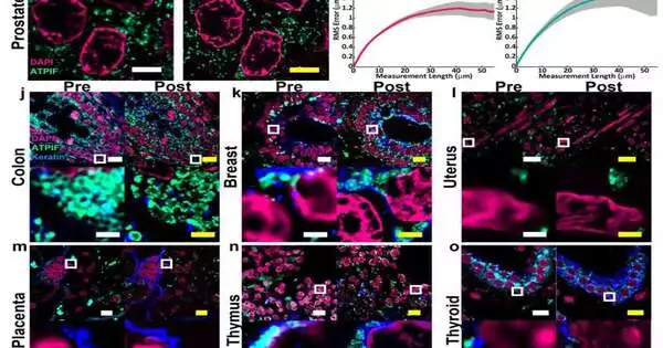
For specialists, for example, Simon Watkins, the pioneer and head of the Middle for Biologic Imaging at the College of Pittsburgh and the Pittsburgh Disease Organization, the way that the new convention is viable with a wide scope of tissue types—iincluding protected tissue segments—iis significant. For instance, most extension microscopy techniques are streamlined for brain tissue. Conversely, Amplify was tested on examples from different human organs, comparing cancers including those of the bosom, mind, and colon.
“Suppose you have a tissue with thick and non-thick parts; this gets around tissues that beforehand wouldn’t extend isometrically,” Watkins said. “Leon has been buckling down on this to make this convention work with tissues that have been filed.”
Xi (Charlie) Ren, an associate teacher of biomedical design at Carnegie Mellon, concentrates on the lung tissue and how to show its morphogenesis and pathogenesis. Some of his research involves looking into the motile cilia that have the ability to clear bodily fluid in the human piloting the aircraft.The designs are too small to see without time-escalated innovation such as electron microscopy, measuring 200 nanometers in width and only a few micrometers in length.Working in a joint effort with Zhao’s lab, Ren’s group created and conveyed lung organoid models with explicit deformities in cilia ultrastructure and worked to approve the capacity of Amplify to imagine clinically important cilia pathology.
“With the most recent Amplify methods, we can extend those lung tissues and begin to see some ultrastructure of the motile cilia even with a standard magnifying instrument, and this will speed up both essential and clinical examinations,” he said.
The analysts likewise had the option to see surrenders in cilia in quiet, unambiguous lung cells known to have hereditary changes.
“The lung tissue design local area in every case needs a superior method for portraying the tissue framework that we work with,” Ren said. He added that this work is a significant initial step, and he trusts that the cooperative work with Zhao’s lab will additionally be refined and applied to pathology tests found in tissue banks.
At long last, the hydrogel utilized in Amplify and created in the Zhao lab is more vigorous than its ancestor, which was exceptionally delicate, causing breaks during the cycle.
“We are wanting to foster this innovation to make it more open to the local area,” he said. “There are various headings this can have.” “There’s a great deal of interest in involving this sort of tissue development innovation in fundamental science.”
Alison Barth, the Maxwell H. and Gloria C. Connan Teacher in the Existence Sciences at Carnegie Mellon, concentrates on the synaptic network during learning. She said the expansive applications given by the new strategies will be an aid for scientists.
“The mind is an incredible spot to exploit these super-goal methods,” said Barth, who teams up with the Zhao Lab on a few examinations. “Microscopy techniques will be useful for synaptic phenotyping and examination across various cerebrum conditions.”
“One of the significant advances in this paper is the strategy’s capacity to chip away at various kinds of tissue examples.”
Extra review creators incorporate Piyumi Wijesekara, Emma F. DiBernardo, and Zhangyu Cheng of Carnegie Mellon; Sinda Fekir and Christopher I. Moore of Earthy Colored College; Donna B. Stolz of Pitt; Franca Cambi of Pitt and the Veterans Organization; and Steven L. Brody and Amjad Horani of Washington College.
More information: Yongxin Zhao, Magnify is a universal molecular anchoring strategy for expansion microscopy, Nature Biotechnology (2023). DOI: 10.1038/s41587-022-01546-1. www.nature.com/articles/s41587-022-01546-1


