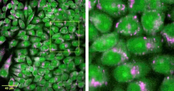
Scientists have developed a Raman magnifying lens that can get data many times quicker than a regular Raman magnifying lens. Raman microscopy is a strong, harmless device for performing complex compound examinations of cells and tissues, and this innovation improvement could assist with growing its value in biomedical applications.
“Our high-throughput Raman ghastly imaging can rapidly picture and examine a huge region with no example pretreatment, which could make it helpful for clinical findings and the tests used to evaluate new medications,” said research group pioneer Katsumasa Fujita from Osaka College. “The strategy’s nameless, high-throughput multiplex compound imaging and examination could also be used to enable new applications or overcome limitations of current techniques.”
In the Optica Distributing Gathering diary, published by Biomedical Optics Express, the analysts describe their new multiline light confocal Raman microscopy approach. It works by recognizing separate areas of the example in equal measure, empowering quick Raman hyperspectral imaging. They demonstrate the way that the method can get hyperspectral pictures of organic tissue with a field of perspective of 1380 x 800 pixels in around 11 minutes. With a standard Raman magnifying lens, this would take days.
“We trust that high-throughput Raman imaging will ultimately make it conceivable to carry out clinical findings more effectively and precisely while perhaps empowering analyses that were absurd previously,” said Fujita. “Name-free atomic examination with Raman imaging would likewise be helpful for effectively recognizing drug reactions in cells, supporting medication advancement.”
Capturing compound data more quickly
Raman spectroscopy, which uses light to energize subatomic vibrations, provides significant insights into the compound cosmetics of an example. The subsequent sub-atomic vibrations make a kind of unique compound mark that can be utilized to recognize the example’s piece. Raman microscopy makes this one stride further by getting high-goal ghastly pictures, which are helpful for imaging cells and tissues. Nonetheless, because of the tradeoff between ghostly goal and imaging speed, Raman microscopy hasn’t been viable for use in the center.
The new multiline light methodology expands upon a strategy the examination group recently created known as “line-brightening Raman microscopy.” That method was faster than standard confocal Raman microscopy and allowed for unique imaging of living cells, but it was still too slow for the large area imaging commonly expected for clinical findings and tissue examination.
“To resolve this issue, we created multiline light Raman microscopy, which gets huge region pictures multiple times quicker than line-brightening Raman microscopy,” said Fujita. “With our new method, the ghastly pixel numberigor goalanand imaging pace can be changed, contingent upon the application. Later on, a much quicker imaging rate may be conceivable as cameras keep on being created with additional pixels.
Gathering the framework
The group’s new multiline-light Raman magnifying lens illuminates around 20,000 focuses in an example, all the while emitting various line-formed laser rays. The Raman dispersing spectra created from the lit positions are then kept in a solitary openness that contains the spatial data for the Raman spectra in the example. Checking how the laser radiates across the example permits a two-layered hyperspectral Raman picture to be recreated.
To accomplish this, the researchers use a round and hollow focal point cluster—an optical component made up of periodically adjusted various tube-shaped focal points—to generate a large number of line-like laser rays from a single laser bar.They combined this with a spectrophotometer capable of acquiring 20,000 spectra at the same time.Optical channels were also important for avoiding cross talk between spectra at the spectrophotometer finder.
A high-responsive, low-clamor CCD camera with countless pixels was likewise basic. “This CCD camera permitted 20,000 Raman spectra to be conveyed on the CCD chip and identified all the while,” said Fujita. “The hand-crafted spectrophotometer likewise assumed a significant role by shaping the 2D dispersion of spectra on the camera without huge bending.”
Testing execution
The analysts utilized the new method to get estimations from live cells and tissues to test its imaging execution and likely use in biomedical applications. They showed that lighting a mouse mind test with 21 concurrent brightening lines could be utilized to get 1,108,800 spectra in 11.4 minutes. They also estimated mouse kidney and liver tissue and led name-free live-cell atomic imaging.
“Little particle imaging and super-multiplex imaging utilizing Raman labels and tests could likewise profit from this method since they don’t need countless pixels in a range and can benefit from quick imaging,” said Fujita.
For this method to be applied for clinical findings, the scientists say it would be vital to fabricate a data set of Raman pictures, something that can be achieved effectively with the new Raman magnifying lens because of its speed and huge imaging region. They are likewise attempting to speed up by an element of around 10 and might want to lessen the expense of the camera, laser, and spectrophotometer to make commercialization more viable.
More information: Kentaro Mochizuki et al, High-throughput line-illumination Raman microscopy with multislit detection, Biomedical Optics Express (2023). DOI: 10.1364/BOE.480611


