Diagnostic imaging is essential in healthcare because it allows doctors to detect and diagnose a variety of medical conditions. However, despite significant advances in imaging technology, existing single imaging modalities are insufficient to cover all diagnostic scenarios, leading to reliance on multiple imaging modalities and increasing healthcare costs. In response to these challenges, researchers at the University of Illinois at Urbana-Champaign have developed a dual-modality imaging technology that not only provides comprehensive diagnostic information but also provides a cost-effective solution for healthcare providers.
Ultrasound imaging (US) is a common and widely used diagnostic tool in the medical field. However, it is limited to low image quality and often needs to be accompanied by higher-quality and more expensive imaging modalities such as MRI. To improve US imaging, Department of Electrical and Computer Engineering assistant professors Chen Yun-Sheng and Yang Zhao, along with ECE graduate student Shengsheng Zhao, combine optoacoustic (PA) imaging, high-resolution ultrasound imaging, and limited deficits. An optimization method for dual high-resolution medical imaging. The new study, titled “Hybrid Optoacoustics and Fast Super-Resolution Ultrasound Imaging,” was recently published in the journal Nature Communications.
“The ability to offer multi-dimensional information is this imaging’s key advantage. We can add various imaging layers. Two layers may be present: a structural layer and a functional one.”
Assistant professors Yun-Sheng Chen .
Zhao said this new imaging tool “improves accessibility, portability, and cost-effectiveness. This method offers similar capabilities and is significantly less expensive than clinical medical imaging methods.”
Traditional imaging methods are most effective in identifying the disease when the patient presents structural changes in the organ. Unfortunately, the disease is often at an advanced stage when these changes are evident. To overcome these drawbacks, the researchers wanted to develop a comprehensive imaging technique capable of detecting additional physiological and biochemical abnormalities, such as changes in blood flow and tissue oxygenation. By combining two new ultrasound imaging technologies, the team hopes to facilitate early disease detection and improve patient outcomes and prognosis. Photoacoustic imaging is a technology that uses laser pulses to generate ultrasound signals that are absorbed by body tissues to create images. Interestingly, when exposed to light, each tissue exhibits a unique signature that allows both tissue structure and content to be identified. PA imaging can image physiological and biochemical processes in the body, such as blood oxygen levels, tissue composition, inflammation, and the distribution of imaging agents (functional information).
Despite the advantages of PA imaging, it also has limitations. Due to its limited resolution, it is not possible to analyze microstructures such as micro vessels, which are important targets in several diseases, including cancer and kidney diseases. High-resolution ultrasound imaging uses microbubbles as contrast agents that flow in the blood and emit very strong ultrasound signals. Using advanced signal processing, ultrasound removes background signals from the tissue and focuses only on the signals from individual imaging agents in the blood.
This technology can achieve high resolution, making it ideal for microvascular imaging and blood flow measurements. Both high-resolution ultrasound imaging and optoacoustic imaging can share the same ultrasound imaging system, making it ideal for dual-modality imaging development. Combining high-resolution US images with PA images may seem straightforward, but each technique has hardware and acquisition speed requirements. The variable recording rate is particularly important because the displayed information can change over time due to the patient’s natural movements, such as breathing and heart rate. Applying constrained optimization to signal processing equalizes the speed of the two imaging techniques and enables smooth scanning of the image area. This approach allowed us to accelerate the frame rate of high-resolution ultrasound images by a factor of 37 for synthetic data and by a factor of 28 for in vivo data. Matching frame rates allow continuous recording of both types of images, enabling successful co-registration of chemical composition, blood flow, and structure.
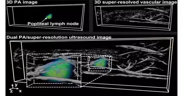
The research team demonstrated the innovative technology in two important in vivo scenarios: lymph nodes and kidneys. The lymph nodes are one of the first places where cancer spreads, and doctors will examine a person’s lymph nodes to determine if a lymphadenectomy is necessary. During this process, it is important to know where the lymph nodes are and to visualize the complex blood vessels around the cancer cells.
Current technology allows you to see blood vessels or lymph nodes, but not both. It has been shown that this new method can provide information about the location of both lymph nodes and blood vessels. The kidneys are another major target, as about 15 percent of Americans have kidney disease, and 40 percent of people with chronic kidney disease are unaware of their condition until diagnostic imaging is performed. Blood vessels and oxygen supply to the kidneys play an important role in the progression of chronic kidney disease. However, as with lymph nodes, current imaging techniques do not equally capture structural and functional information.
The interactive imaging method allows the determination of specific blood vessels in the kidney as well as the function of the kidney tissue. Chen: “The great thing about this image is that it can provide multidimensional information. You can add other layers to the image. One layer can be structural, and another layer can be functional.” Next, they want to add another layer, molecular information, to track changes at the molecular or genetic level, where most diseases occur.
The team, along with other campus staff members, are excited about the opportunity to use this new imaging method. They are interested in using the method to study neurodegenerative diseases of the brain. Given the critical role of oxygen delivery in brain function, this imaging technique could be a powerful tool for imaging the vasculature that supplies brain oxygen.
More information: Shensheng Zhao et al, Hybrid photoacoustic and fast super-resolution ultrasound imaging, Nature Communications (2023). DOI: 10.1038/s41467-023-37680-w


