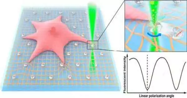
Mechanics plays a key role in cell science. Cells explore these mechanical powers to investigate their surroundings and sense the way of behaving encompassing living cells. The actual qualities of a cell’s current circumstances thus influence the cell’s capabilities. Understanding how cells interact with their current surroundings provides valuable experience in cell science and has broader implications in medicine, including disease detection and treatment.
Up to this point, analysts have fostered various devices to concentrate on the exchange among cells and their 3D microenvironment. One of the most famous advances is footing force microscopy (TFM). It is a main strategy to decide the footings on the substrate surface of a cell, giving significant data on how cells sense, adjust and respond to the powers.
In any case, TFM’s application is restricted to giving data on the translational movement of markers on cell substrates. Data about different levels of opportunity, for example, rotational movement, stays speculative because of specialized limitations and restricted research on the point.
“The majority of cells in multicellular organisms are subjected to forces that are highly coordinated in space and time, and developing a multi-dimensional cell traction force field microscope has been one of the field’s most difficult problems.”
Dr. Zhiqin Chu of the Department of Electrical and Electronic Engineering
Design specialists at the College of Hong Kong have proposed a clever method to gauge the cell footing force field and tackle the examination hole. The interdisciplinary exploration group was led by Dr. Zhiqin Chu of the Branch of Electrical and Electronic Design and Dr. Yuan Lin of the Branch of Mechanical Design. They utilized single nitrogen-opening (NV) focuses in nanodiamonds (NDs) to propose a direct polarization tweak (LPM) strategy which can gauge both the rotational and translational development of markers on cell substrates.
The review gives another viewpoint on the estimation of complex cell footing force fields and the outcomes have been published in the journal Nano Letters.
The examination showed high-accuracy estimations of rotational and translational movement of the markers on the cell substrate surface. These trial results prove the hypothesis and past outcomes.
Given their ultrahigh photostability, great biocompatibility, and helpful surface compound change, fluorescent NDs with NV focuses are superb fluorescent markers for the majority of organic applications. The scientists found that in view of the estimation consequences of the connection between the fluorescence power and the direction of a solitary NV point to laser polarization course, high-accuracy direction estimations and foundation free imaging could be accomplished.
Hence, the LPM strategy created by the group settles specialized bottlenecks in cell force estimation in mechanobiology, which envelops interdisciplinary joint efforts from science, design, and physical science.
“Most cells in multicellular creatures experience powers that are profoundly arranged in reality. The improvement of a complex cell footing force field microscopy has been perhaps the best test in the field, “said Dr. Chu.
“Contrasted with the regular TFM, this new innovation furnishes us with a new and helpful device to examine the genuine 3D cell-extracellular grid connection. It accomplishes both quantitative and qualitative translational development estimations in the cell footing field and uncovers data about the cell foothold force, “he added.
The review’s primary feature is the capacity to show both the translational and rotational movement of markers with high accuracy. It is a major step towards examining mechanical connections at the cell-grid interface. It, likewise, offers new roads of exploration.
Through specific synthetics on the phone surface, cells interface and interface as a feature of a cycle called cell grip. The manner in which a phone creates strain during bond has been basically depicted as’ in-plane.’ For example, footing pressure, actin stream, and grip development are totally associated and show complex directional elements.
The LPM strategy could assist with figuring out the muddled forces encompassing central grip and separating different mechanical burdens at a nanoscale level (e.g., typical footings, shear powers). It might likewise assist with understanding how cell bonds respond to various sorts of pressure and how these intervene in mechanotransduction (the system through which cells convert mechanical boost into electrochemical action).
This innovation is also encouraging research into various other biomechanical processes, such as safe cell enactment, tissue arrangement, and the replication and attack of disease cells. For instance, lymphocyte receptors, which play a focal part in safe reactions to disease, can create very unique powers crucial to tissue development. This high-accuracy LPM innovation might assist with examining these complex power elements and give experience in tissue advancement.
The examination group is effectively exploring systems to grow optical imaging abilities and, all the while, map various Nano diamonds.
More information: Lingzhi Wang et al, All-Optical Modulation of Single Defects in Nanodiamonds: Revealing Rotational and Translational Motions in Cell Traction Force Fields, Nano Letters (2022). DOI: 10.1021/acs.nanolett.2c02232
Journal information: Nano Letters


