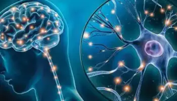
Researchers may now investigate the gene expression profiles of individual brain cells and obtain insights into the diversity and function of distinct cell types in the brain thanks to advances in single-cell sequencing and imaging technologies, as well as techniques like single-cell RNA sequencing. These technologies have revealed important details on brain cell heterogeneity and their roles in numerous neurological processes.
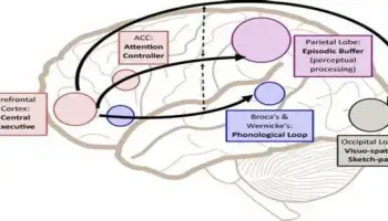
Researchers generated a detailed cellular map of Broca’s area in the human brain by combining non-invasive imaging techniques.
The brain is made up of various cell types that are organized into various structures and regions. Although significant progress has been made toward building human brain models, the breakthroughs have not yielded undistorted 3D representations of cellular architecture, which are required to build precise and detailed models.
A team led by investigators at Massachusetts General Hospital (MGH), a founding member of Mass General Brigham (MGB), has overcome this challenge in new research published in Science Advances to offer scientists and clinicians a comprehensive cellular atlas of a part of the human brain known as Broca’s area, with a detailed resolution to study brain function and health.
We built the technology needed to integrate information across many orders of magnitude in spatial scale from images in which pixels are a few microns to those that image the entire brain.
Bruce Fischl
The researchers were able to create a high-resolution cell census atlas of a specific region of the human cerebral cortex, or the outer layer of the brain’s surface, by combining different sophisticated imaging techniques, including magnetic resonance imaging, optical coherence tomography, and light-sheet fluorescence microscopy.
The scientists developed such an atlas for a human postmortem specimen and integrated it into a whole-brain reference atlas.
“We built the technology needed to integrate information across many orders of magnitude in spatial scale from images in which pixels are a few microns to those that image the entire brain,” says co-senior author Bruce Fischl, Ph.D., director of the Computational Core at the Athinoula A. Martinos Center for Biomedical Imaging at MGH and a professor in Radiology at Harvard Medical School.
Ultimately, the methods in this study could be used to reconstruct undistorted 3D cellular models of particular brain areas as well as of the whole human brain, enabling investigators to assess variability between individuals and within a single individual over time.
This will provide insights into the presence and spread of pathologic changes that occur in neurodegenerative and psychiatric illnesses.
“These breakthroughs will help us understand the mesoscopic structure of the human brain, which we currently know very little about.” Structures that are too vast and geometrically complicated to be studied by looking at 2D slices on a conventional microscope stage, yet too small to observe routinely in living human brains, according to Fischl.
“Currently we don’t have rigorous normative standards for brain structure at this spatial scale, making it difficult to quantify the effects of disorders that may impact it such as epilepsy, autism, and Alzheimer’s disease.”


