Because X-ray microscopes can penetrate most materials, chest X-rays and CT scans allow non-invasive examination of internal organs and skeletons. In recent times, efforts have been made to improve the resolution of X-ray imaging technology in order to precisely observe the internal structure of batteries and semiconductors at the nanoscale.
A core technology that can overcome the resolution limitations of existing X-ray microscopes has been developed by a joint research team led by Professor YongKeun Park of the KAIST Department of Physics and Dr. Jun Lim of the Pohang Accelerator Laboratory.
“The resolution in this study was limited to 14 nm, but if the next-generation X-ray light source and high-performance X-ray detector are used, the resolution would exceed that of conventional X-ray nano-imaging and approach the resolution of an electron microscope.”
Dr. KyeoReh Lee of KAIST Department of Physics,
Dr. KyeoReh Lee was the first author of this study, which was published in Light on April 6: Science and How It Can Be Used. “Direct high-resolution X-ray imaging exploiting pseudorandomness” is the title of it.
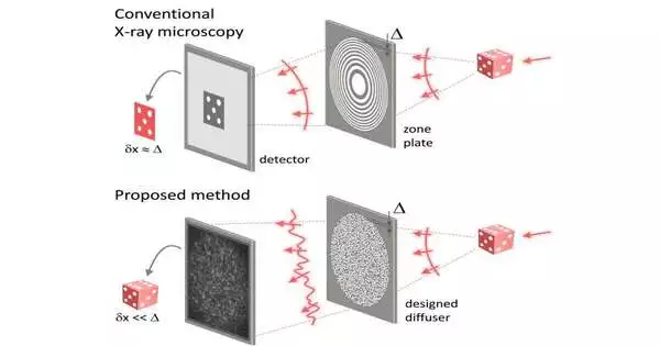
Refractive lenses are not present in X-ray nanomicroscopes. A concentric zone plate, a circular grating, replaces the lens in an X-ray microscope. The quality of the plate’s nanostructure determines the resolution of an image obtained with the zone plate. The level of resolution that can be achieved with X-ray microscopy is constrained by a number of issues that arise during the process of creating and maintaining these nanostructures.
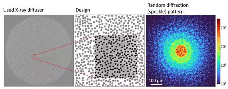
Fig. 2. A surface electron microscopy (SEM) image of the experiment’s X-ray diffuser can be seen in the left panel. The center board shows the plan of the X-beam diffuser, and there is an inset in the board that shows a comparison of some portions of the SEM picture. The right board shows an exploratory irregular X-beam diffraction design, otherwise called a spot design, obtained from the X-beam diffuser. Credit: The Korea Advanced Institute of Science and Technology (KAIST).
A new method for X-ray nanomicroscopy to solve this issue. By diffracting incident X-rays, the team’s X-ray lens, which takes the form of numerous holes punched into a thin tungsten film, generates random diffraction patterns. Mathematically, the researchers were able to determine that, paradoxically, the sample’s high-resolution information was completely contained in these random diffraction patterns. They were also successful in extracting the information and imaging the samples’ internal states.
In 2016, Dr. KyeoReh Lee and Professor YongKeun Park proposed and first implemented an imaging technique in the visible light band based on the mathematical properties of random diffraction. This study utilizes the aftereffects of past examinations to address the troublesome, waiting issue in the field of ble light band based on the mathematical properties of random diffraction. This study utilizes the aftereffects of past examinations to address the troublesome, waiting issue in the field of X-ray imaging.
There is no direct correlation between the size of the pattern etched on the random lens and the resolution of the image of the constructed sample. Using random lenses that were made in a circular pattern and had a diameter of 300 nm, the research team was able to obtain images with a resolution of 14 nm (approximately 1/7 the size of the coronavirus). This idea served as the foundation for their findings.
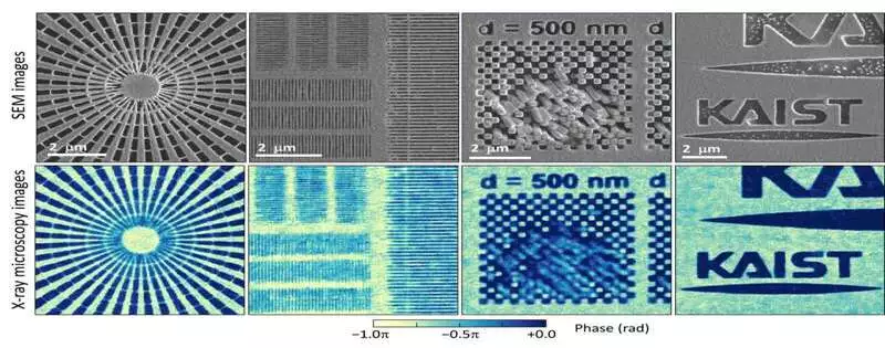
Fig. 3. Surface electron microscope (SEM) images taken in conjunction with the proposed randomness-based X-ray imaging (bottom) Credit: The Korea Advanced Institute of Science and Technology (KAIST) .
This research team’s imaging technology is a crucial, fundamental technology that has the potential to improve the resolution of X-ray nanomicroscopy but has been hindered by the production limitations of the existing zone plates.
“In this study, the resolution was limited to 14 nm, but if the next-generation X-ray light source and high-performance X-ray detector are used, the resolution would exceed that of conventional X-ray nano-imaging and approach the resolution of an electron microscope,” stated Dr. KyeoReh Lee of the KAIST Department of Physics, one of the co-corresponding authors.
“X-rays will be able to present a new standard for non-invasive nanostructure observation processes such as quality inspections for semiconductors because, unlike an electron microscope, they can observe the internal structure without damaging the sample,” the statement reads.
“In the same context, the developed image technology is expected to greatly increase the performance of the 4th generation multipurpose radiation accelerator, which is set to be established in Ochang, Northern Chungcheong Province,” stated Dr. Jun Lim, co-corresponding author from the Pohang Accelerator Laboratory.
More information: KyeoReh Lee et al, Direct high-resolution X-ray imaging exploiting pseudorandomness, Light: Science & Applications (2023). DOI: 10.1038/s41377-023-01124-3


