To make due, creatures should control the strain inside them, from the single-cell level to tissues and organs. Estimating these tensions in living cells and tissues in physiological circumstances is a test.
In research that has its starting point at UC St. Nick Barbara, researchers now at the Group of Greatness Physical Science of Life (PoL) at the Specialized College in Dresden (TU Dresden), Germany, report in the diary Nature Correspondences another method to ‘picture’ these tensions as creatures create. These estimations can assist with understanding how cells and tissues get by under tension and uncover how issues in controlling tensions lead to sickness.
At the point when particles break down in water and are isolated into various compartments, water tends to move, starting with one compartment and then onto the next, to equilibrate their focuses, a cycle known as assimilation. In the event that a few particles can’t cross the layer that isolates them, a tension irregularity—osmotic strain—develops between compartments.
“We know that various physical mechanisms influence how our bodies function. Osmotic pressure, in particular, is recognized to play a critical role in the development of organs during embryogenesis, as well as in the maintenance of healthy adult organs. We can now analyze how osmotic pressure affects all of these processes directly in real tissues using this new approach.”
Led by former UCSB professor Otger Campàs,
This guideline is the reason for the majority of specialized applications, for example, the desalination of seawater or the improvement of saturating creams. It would seem that keeping a sound, working life makes the rundown as well.
Our cells are continually moving atoms in and out to prevent the strain that develops from pounding them. To do so, they utilize atomic siphons that permit them to hold the tension in line. This osmotic strain influences numerous parts of cells’ lives and even sets their size.
At the point when cells collaborate to fabricate our tissues and organs, they, too, face a tension issue: Our vascular framework, or organs like the pancreas or liver, contain liquid-filled pits known as lumens that are fundamental for their capability. Assuming cells neglect to control osmotic tension, these lumens might implode or detonate, with possibly devastating ramifications for the organ.
To comprehend how cells control strain in these tissues, or how they neglect to do so in illness, it is crucial to measure and ‘see’ the osmotic tension in live tissues. In any case, sadly, this was impractical.
As of not long ago.
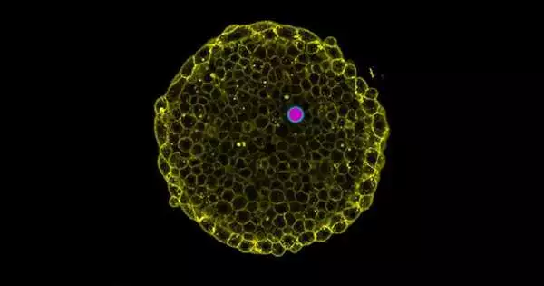
Driven by previous UCSB teacher Otger Campàs, who presently holds the Seat of Tissue Elements at TU Dresden and is as of now the overseeing head of PoL, the researchers formulated an original procedure to quantify the osmotic strain in living cells and tissues by utilizing exceptional beads known as twofold emulsions.
For this strain sensor, they brought a water drop into an oil drop that allowed water to move through. When these “twofold drops” were presented to salt arrangements of various focuses, water streamed all through the inside water bead, changing its volume, until pressures were equilibrated. The specialists demonstrated the way that the osmotic tension can be estimated by just checking the drop size. They then brought these twofold beads into living cells and tissues, utilizing glass microcapillaries to uncover their osmotic strain.
“It just so happens that cells in creature tissues have similar osmotic tension as plant cells. Be that as it may, dissimilar to plants, they should offset it continually with their current circumstances to abstain from detonating since they don’t have unbending cell walls,” Campàs said.
With this straightforward idea, this shrewd technique currently permits researchers to “see” osmotic strain in a large number of settings. “We realize that few actual cycles influence the working of our bodies,” Campàs said. “Specifically, osmotic tension is known to assume a central role in the structure of organs during embryogenesis and, furthermore, in the upkeep of solid grown-up organs. With this new strategy, we can currently concentrate on what osmotic strain means for this large number of cycles straightforwardly in living tissues.”
Past contribution experiences into the natural cycles and actual rules that oversee life, this technique holds promising modern and clinical applications, including observing skin hydration, portrayal of creams or food varieties, and determination of sicknesses known to have osmotic strain-lopsided characteristics, like cardiovascular infections or growths. The patent for this method is as of now being given by UC St. Nick Barbara, where Campàs carried out his analysis prior to joining TU Dresden.
Campàs’ lab recently created interesting procedures to gauge the small powers that cells make inside tissues and, furthermore, extra actual properties utilizing infinitesimal single beads. Antoine Vian, the lead creator of the work and a specialist in microfluidics, the innovation that empowers the age of twofold emulsion beads, accentuated their key job.
“Twofold emulsions are extremely adaptable, with a wide range of applications in science and innovation,” he said. “Single drops can be distorted; however, they are incompressible and don’t permit pressure estimations. Interestingly, twofold emulsion drops can change size and be utilized as osmotic strain sensors. Their utilization in living frameworks will doubtlessly empower previously unheard-of revelations.”
More information: Antoine Vian et al, In situ quantification of osmotic pressure within living embryonic tissues, Nature Communications (2023). DOI: 10.1038/s41467-023-42024-9


