Scientists at Nagoya College in Japan have utilized another gadget to recognize a key protein in pee that shows whether the patient has cerebrum cancer. This protein could be used to detect cerebrum malignant growth, eliminating the need for invasive tests and increasing the likelihood of cancers being detected early enough for treatment. This examination could likewise have had ramifications for recognizing different sorts of malignant growth. The exploration was distributed by ACS Nano.
Albeit early recognition of many sorts of malignant growth has added to the expansion of disease endurance rates, the endurance rate for cerebrum cancers has remained practically unaltered for more than 20 years. Incompletely, this is because of their late location. Doctors frequently find cerebrum growths solely after the beginning of neurological side effects, like loss of development or discourse, by which time the cancer has arrived at an impressive size. Identifying the cancer when it is still small and beginning therapy as quickly as time permits ought to assist in saving lives.
One potential sign that an individual has mind cancer is the presence of growth-related extracellular vesicles (EVs) in their pee. EVs are nanosized vesicles engaged in various capabilities, including cell-to-cell correspondence. Since those found in the minds of malignant growth patients have explicit sorts of RNA and film proteins, they could be utilized to recognize the presence of disease and its movement.
“Many body fluids can be used for liquid biopsy, although blood tests are invasive. Because urine includes many useful biomolecules that may be traced back to diagnose the disease, urine tests are an effective, simple, and non-invasive approach.”
Associate Professor Takao Yasui of Nagoya University Graduate School of Engineering.
In spite of the fact that they are discharged a long way from the mind, numerous EVs from malignant growth cells exist steadily and are discharged in the urine without separating. Peer testing enjoys many benefits, which makes sense to academic administrator Takao Yasui of Nagoya College’s Graduate School of Design. “Fluid biopsies can be performed utilizing many body liquids, yet blood tests are intrusive,” he said. “Pee tests are a powerful, straightforward, and harmless technique in light of the fact that the pee contains numerous enlightening biomolecules that can be followed back to distinguish the sickness.”
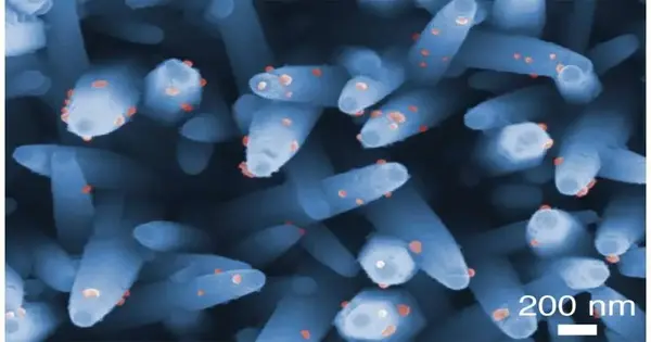
An examination bunch led by Yasui and Teacher Yoshinobu Baba of Nagoya College’s doctoral-level college of designing, as a team with Nagoya College’s Organization of Advancement for Future Society and the College of Tokyo, has fostered another investigation stage for mind cancer EVs utilizing nanowires at the lower part of a well plate. Using this device, they identified two distinct types of EV layer proteins, known as CD31 and CD63, in urine samples from brain-growth patients.Searching for these obvious proteins could empower specialists to recognize cancer patients before they foster side effects.
“Presently, EV detachment and identification strategies require multiple instruments and a measure to segregate and then distinguish EVs,” said Yasui. “With a single simple strategy, the overall nanowire examination can confine and identify EVs.” Later on, clients can run tests through our examine and change the location part by specifically adjusting it to distinguish explicit film proteins or miRNAs inside EVs to recognize different sorts of malignant growth. Utilizing this stage, we hope to propel the examination of the articulation levels of explicit film proteins in patients’ urinary EVs, which will empower the early identification of various kinds of disease.
More information: Kunanon Chattrairat et al, All-in-One Nanowire Assay System for Capture and Analysis of Extracellular Vesicles from an ex Vivo Brain Tumor Model, ACS Nano (2023). DOI: 10.1021/acsnano.2c08526


