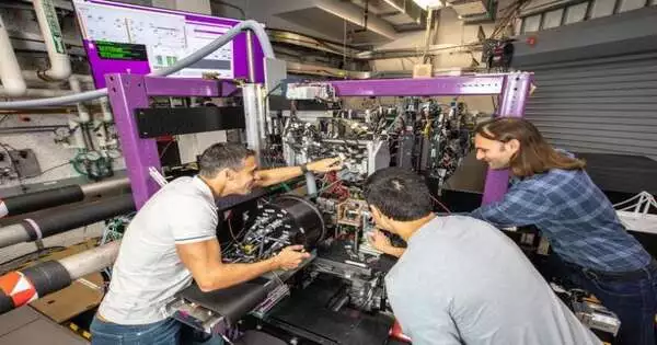
By combining two ultrafast X-ray spectroscopy methods, researchers at SLAC National Accelerator Laboratory were able to record for the first time one of ferricyanide’s fastest movements. They figure their methodology could assist with planning more perplexing compound responses like oxygen transportation in platelets or hydrogen creation utilizing fake photosynthesis.
The research team from Stanford, SLAC, and other institutions started with a method that is now fairly common: they used an ultraviolet laser and the Linac Coherent Light Source (LCLS) X-ray free-electron laser to zap a mixture of ferricyanide and water. The bright light kicked the particle into an invigorated state while the X-beams tested the example’s molecules, uncovering highlights of ferricyanide’s nuclear and electronic design and movement.
What was different this time was the way the specialists removed data from the X-beam information. The team captured and examined valence-to-core, a second emission region that has been significantly more difficult to measure on ultrafast timescales, as opposed to only one spectroscopic region, the K main emission line. The team was able to get a comprehensive picture of the ferricyanide molecule as it evolved into a crucial transitional state by combining data from both areas.
“There was no specific experimental evidence for each of the various phases in the basic chemical reaction known to occur in ferricyanide, which is the ligand exchange.”
SLAC scientist and first author Marco Reinhard
The group showed that ferricyanide enters a middle, invigorated state for around 0.3 picoseconds — or under a trillionth of a second — subsequent to being hit with an UV laser. The valence-to-core measurements then showed that ferricyanide loses one of its molecular cyanide “arms,” or ligands, following this brief excited period. The missing joint is then either filled with the same carbon-based ligand or, less likely, with a water molecule by ferricyanide.
“This ligand trade is an essential synthetic response that was remembered to happen in ferricyanide; however, there was no immediate trial proof of the singular strides in this cycle,” SLAC researcher and first creator Marco Reinhard said. “We wouldn’t really be able to see how the molecule changes from one state to the next using only a K main emission line analysis method; we would only have a clear picture of the process’s beginning.”
“You need to have the option to reproduce how nature further develops innovation and increments our primary logical information,” SLAC senior researcher Dimosthenis Sokaras said. “In addition, you need to be familiar with each step, from the most obvious to the less obvious, so to speak, in order to replicate natural processes more accurately.”
Later on, the examination group needs to concentrate on additional mind-boggling particles, for example, hemeproteins, which transport and store oxygen in red platelets —which can be precarious to study since researchers don’t see every one of the middle-of-the road steps of their responses, Sokaras said.
Over the course of many years, the research team improved their X-ray spectroscopy method at SLAC’s Stanford Synchrotron Radiation Lightsource (SSRL) and LCLS, combining this knowledge with the X-ray Correlation Spectroscopy (XCS) instrument at LCLS to capture ferricyanide’s molecular structural changes. The group distributed their outcomes today in Nature Correspondences.
“To finish the experiment, we used both SSRL and LCLS. We could never have wrapped up fostering our technique without admittance to the two offices and our longstanding coordinated effort,” said Roberto Alonso-Mori, SLAC lead researcher. “At these two X-ray sources, we have been developing these techniques for years, and we now intend to use them to uncover previously unknown chemical reaction secrets.”
More information: Marco Reinhard et al, Ferricyanide photo-aquation pathway revealed by combined femtosecond Kβ main line and valence-to-core x-ray emission spectroscopy, Nature Communications (2023). DOI: 10.1038/s41467-023-37922-x


