A group of scientists at McGill College, working with an associate from Charité-Universitätsmedizin in Germany, has uncovered a piece of the cycle utilized by mussels to tie to rocks and to let out of them when conditions warrant it.
In their venture, announced in the journal Science, the gathering concentrated on the connection point between mussel tissue and the heap of fibers that mussels use to secure themselves to rocks and different articles. Guoqing Container and Canister Li, along with Jiangsu College and Soochow College, both in China, have distributed a point-of-view article in a similar diary issue framing the work done by the group on this new exertion.
Mussels are bivalve mollusks that live in both freshwater and saltwater conditions. They have pivoted shells that are joined by a tendon. Muscles guarantee a tight seal when the shell is shut. Mussels use byssus strings (referred to ordinarily as facial hair growth) to join themselves to strong items like rocks.
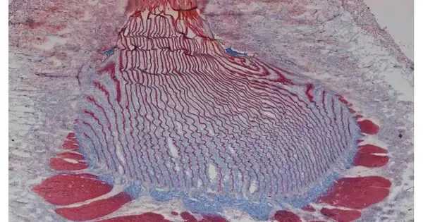
3D remaking of the miniature registered tomography (CT) dataset This film was produced from the recreation of a μCT dataset gained from a f stem root stained implanted in living tissue. At first, the generator and septa tissues are pictured in grayscale, while the stem root lamellae are envisioned in light blue. Moving from the outer stem area into the stem root, one can see the perplexing interdigitation of the septae and lamellae. Later in the film, the generator tissue is eliminated from the model to all the more clearly envision the complicated design of the wavy lamellar sheets. Credit: Jenaes
Sivasundarampillai
The mussel byssus has been broadly concentrated because of its extraordinary capacity to interface nonliving material (the fibers that make up the strings) with living tissue and to separate on request. In any case, as Skillet and Li note, the greater part of this examination has spun around conceivable compound-restricting components. In this new exercise, the examination group focused more on the elements of the biointerface.
To more readily comprehend how the byssus strings interface with living tissue and how they can be cast off if necessary, the exploration group utilized different advancements to concentrate on the strings and the tissue with which they associate. By utilizing a few kinds of imaging alongside spectroscopy, the group saw that the closures of the strings interlocked with layers of living tissue, which themselves were covered with roughly 6 billion motile cilia.
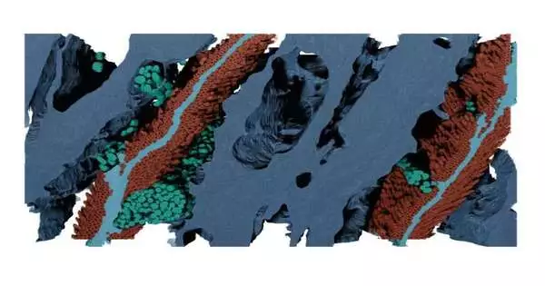
Reproduced highlights in 3D from a Lie SEM picture stack produced using a little district in the stem root Living tissue is in dull blue; non-living stem root sheets are in light blue; secretory vesicles are in greenish blue; and cilia are in red. Credit: Jenaes Sivasundarampillai.
They further found that having such countless cilia meant a serious level of surface contact, which considered precisely fitting two different materials. The specialists additionally noticed that cilia motions served to both reinforce the hold between the two materials and take into account fast delivery when it was required. They observed that cilia development was driven by synapses, which, the scientists estimate, recommends that they are at last constrained by serotonin and dopamine.
More information: Jenaes Sivasundarampillai et al. A strong quick-release biointerface in mussels mediated by serotonergic cilia-based adhesion, Science (2023) DOI: 10.1126/science.adi7401
Guoqing Pan et al, A dynamic biointerface controls mussel adhesion, Science (2023). DOI: 10.1126/science.adl2002


