Metabolic issues assume a focal role in numerous normal circumstances, including Alzheimer’s, the downturn, diabetes, and disease, which call for solid as well as harmless demonstrative methodology. As part of the process of mapping the brain’s glucose metabolism up until this point, radioactive substances have been given to the brain.
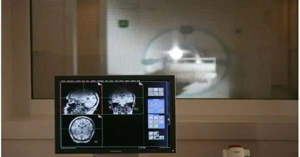
Presently, a MedUni Vienna research group has fostered a totally new, attractive reverberation imaging (X-ray) approach. The procedure, which makes use of a non-toxic glucose solution, yields reliable results and, in theory, is compatible with all common MRI scanners. The study’s findings were recently published in the Nature Biomedical Engineering journal.
The study examined and substantially improved existing diagnostic methods for mapping brain glucose metabolism. During a period of approximately 90 minutes, blood glucose levels and metabolic products were measured multiple times in healthy subjects. Rather than existing methodology, the subjects didn’t get radio-named glucose, but an amount of an innocuous glucose arrangement comparable to a jar of a bubbly beverage. As this substance doesn’t deliver an immediate sign for the MR imaging technique utilized, fixations and digestion of glucose were estimated by implication in light of the drop in signal power for the item concerned.
“The key benefit of this indirect method is that, unlike previous, comparable approaches, no additional hardware components are needed, therefore it can be utilized without any issues on other MR devices.”
Principal investigator Wolfgang Bogner of the Department of Biomedical Imaging and Image-guided Therapy at MedUni Vienna.
“The main advantage of this indirect method is that it can be used on other MR devices without any difficulties because it does not require additional hardware components, as is the case with other, comparable approaches,” said the study’s principal investigator, Wolfgang Bogner, of MedUni Vienna’s Department of Biomedical Imaging and Image-guided Therapy, emphasizing the findings’ clinical significance.
Wide range of potential applications The high-performance 7 Tesla MRI scanner at MedUni Vienna was used in the study, which was carried out by researchers from the Department of Psychiatry and Psychotherapy and the Department of Medicine III. Since its inception in 2008, the device has served as Austria’s sole ultra-high-field MR scanner. Wolfgang Bogner and his group have previously demonstrated that the original methodology additionally deals with 3-Tesla MR scanners.
Lead author Fabian Niess stated, “That was an important step, because 3T MR systems are extremely widespread in clinical applications,” in a follow-up study that was published in Investigative Radiology.
More research is needed to confirm the results. Many common diseases have problems with the metabolism of glucose. It is now realized that disease and growth cells consume far more noteworthy amounts of glucose than ordinary cells — an impact that doctors can benefit from while diagnosing and confining cancers.
This is currently accomplished by utilizing positron emission tomography in conjunction with computed tomography (PET-CT), in which patients are required to receive a small amount of radioactive glucose via injection. Nonetheless, the discoveries should be confirmed in additional examinations before the new, less-obtrusive technique created at MedUni Vienna can be conveyed to patients.
More information: Petr Bednarik et al, 1H magnetic resonance spectroscopic imaging of deuterated glucose and of neurotransmitter metabolism at 7 T in the human brain, Nature Biomedical Engineering (2023). DOI: 10.1038/s41551-023-01035-z
Fabian Niess et al, Noninvasive 3-Dimensional 1H-Magnetic Resonance Spectroscopic Imaging of Human Brain Glucose and Neurotransmitter Metabolism Using Deuterium Labeling at 3T, Investigative Radiology (2023). DOI: 10.1097/RLI.0000000000000953


