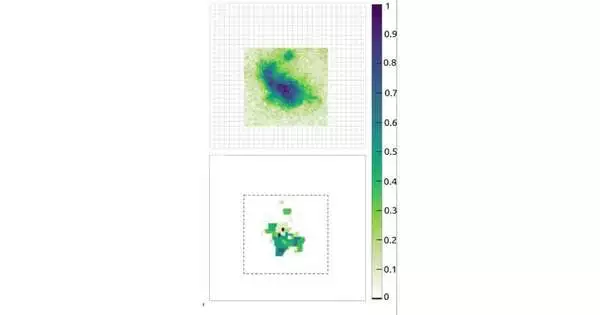
An optical fiber as slim as a strand of hair holds promise for use in negligibly obtrusive profound tissue investigations of patients’ minds that show the impacts of Alzheimer’s illness and other cerebrum issues. The examination could pave the way for negligibly obtrusive in vivo mind imaging in lab studies and check neuronal action over the long run in patients with neurological issues.
“The ultrathin multimode fiber would handily squeeze into a needle therapy needle, and we realize these needles can be embedded into anybody’s body with basically no agony, possibly empowering profound tissue imaging progressively,” said co-creator Benjamin Lochocki, from Vrije Universiteit Amsterdam.
The test is effectively expanding the picture goal at the subcellular level since loss of data is unavoidable from light scrambling. At APL Photonics, analysts in the Netherlands address this test with dot-based compressive imaging (SBCI) that takes advantage of the light scrambling of multimode strands for their potential benefit.
“The ultrathin multimode fiber would easily fit inside an acupuncture needle, and we know these needles can be placed into anyone’s body with almost little pain, possibly enabling real-time deep-tissue imaging,”
Benjamin Lochocki, from Vrije Universiteit Amsterdam.
Optical strands, a sure-known method for guiding light over significant distances, stand out in microendoscopy as a superior method for getting to profoundly lying tissue because of their miniscule aspects. They also eliminate the need for fluorescent naming, which is a cumbersome and costly step.
Light scrambling is commonly tended to through forming the wavefront of an episode bar to lessen dispersing and make an engaged bar at the distal end of the fiber. Nonetheless, this method has limits in securing speed and creating great tissue pictures.
SBCI changes the laser bar passage position to make various uncorrelated irregular dot designs at the fiber yield. A PC calculation can recreate a picture of the item based on the example and its gathered data.
This “compressive imaging” lessens how many pixel estimations are expected to remake a picture of comparable or preferred quality over the best quality level raster imaging utilized in regular endoscopes and magnifying lenses. The SBCIapproach can create high-goal pictures up to multiple times faster, for a space multiple times larger than the customary raster-check approach.
The method was used to image lipofuscin, an age-related fluorescent color that accumulates over time as metabolic waste in the soma, the part of the neurons that contains the core and is in charge of synapse creation.amassing of lipofuscin may be related to Alzheimer’s illness movement, despite the fact that there is minimal understanding of this cycle.
The shade development was imagined in a mind tissue test of an Alzheimer’s patient giver obtained through the Netherlands Brain Bank.
More information: Benjamin Lochocki et al, Epi-fluorescence imaging of the human brain though a multimode fiber, APL Photonics (2022). DOI: 10.1063/5.0080672


