Language, memory, and decision-making are just a few of the higher-order functions that the cerebral cortex, the outer layer of the mammalian brain, is known to be essential for. Numerous studies have looked at its structure and function, but it has been very challenging to image the neuronal dynamics and identify the cell types in behaving animals.
Recently, a study was conducted by scientists at Cold Spring Harbor Laboratory and the Duke University Medical Center with the goal of observing and better comprehending neural circuit dynamics in the cerebral cortex of behaving mice. Their results, which were reported in Nature Neuroscience, demonstrated that while mice engaged in various behaviors, various glutamatergic projection neuron types in the cerebral cortex displayed various patterns of activation.
According to Josh Huang, one of the study’s authors, “the main goal of our study was to understand the neural circuit mechanism underlying brain function, particularly the function of the cerebral cortex.”. “When we ask this, the logical follow-up question is what cell type resolution do we capture, as cells are the fundamental building blocks of neural circuits that form connections and have various functions.”.
“The primary goal of our research was to better understand the neural circuit mechanisms that support brain function, particularly cerebral cortex function.”
Josh Huang, one of the researchers who carried out the study,
Functional magnetic resonance imaging (fMRI), a method that detects and measures minute changes in blood flow related to brain activity, has been used in numerous studies investigating the cerebral cortex up to this point. Despite its usefulness in some contexts, fMRI has a poor ability to record the spatial and temporal resolution of brain activity. As a result, it is not ideal for thoroughly examining neural circuit dynamics.
Wide-field imaging has only recently been used by neuroscientists to examine brain activity. This imaging technique, which uses genetically encoded sensors to more accurately detect changes in brain activity, is promising.
Although wide-field imaging has significantly advanced research, Huang noted that it is typically limited to imaging all neural populations. Again, it does not resolve these fundamental characteristics of neuronal cell types, particularly those of projection neuron types. Our study was driven by the desire to investigate the cerebral cortex using a recently developed systematic set of genetic tools that can resolve various classes and types of glutamatergic projection neurons with cell type resolution.
Huang and colleagues used a variety of genetic and imaging techniques to measure the activity of neurons in the cerebral cortex of mice in their experiments. In order to first ensure that they could precisely track the activity of various projection neuron types, they used genetic engineering.
We targeted projection neurons that project inside the cortex, outside the cortex to subcortical targets, and to the thalamus in this manner, according to Huang. The key in this situation is to screen across cell types by looking at multiple cell types rather than just one. Assuming that this area is what is involved in behavior, we combine this with wide-field imaging to look at multiple areas concurrently rather than just one.
The team’s ability to use wide-field imaging methods to observe various projection neuron types in awake, behaving mice as they engaged in various activities is a significant accomplishment of this study. However, they did this methodically and with better resolution compared to earlier studies, which enabled them to separate cell type function.
We were able to examine the fundamental components of neuron circuits, projection neuron types, and their dynamics in real-time and in behaving animals across the entire cortical network, Huang said, which I believe has never been done before. This improved our understanding of how the cortex is structured by enabling us to identify the cell type basis of cortical network dynamics.”.
Mohan et al.
As of now, it is understood that the cerebral cortex is divided into various regions that each serve a unique purpose. Due to the presence of so-called “canonical” cortical microcircuits, which are vertical clusters of neurons that are arranged similarly, it was discovered that these areas share a similar neuronal circuit organization. Previous research indicates that these circuits communicate and process information with other brain regions that have a similar circuit structure.
According to Huang, “These columns of neurons across various cortical layers are thought of as structural and, in some cases, functional units.”. This opinion has been widely held for a long time and is supported by both anatomical and functional data that has been gathered in the past. To our surprise, we discover that projection neuron types exhibit rather distinctive dynamic patterns when viewed along the vertical axis.”.
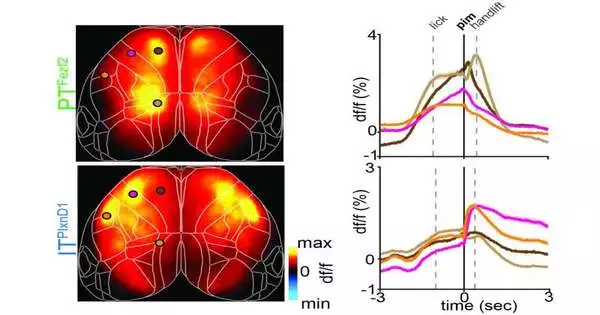
In essence, the intratelencephalic (IT) and pyramidal track (PT) populations of projection neuron types were identified by Huang and colleagues as those that do not always work as a single unit.
Other cortical regions of the cerebral cortex are served by IT neurons. Contrarily, PT neurons transmit data to subcortical parts of the brain outside of the cerebral cortex.
The two major categories of neurons appear to act in different spatial and temporal patterns “in many, but not all, cases,” according to Huang. These observations suggest that these cell types also form distinct processing and output networks, which significantly depart from the more conventional historical concept of columnar neuronal organization.
According to the data gathered by Huang and colleagues, the populations of IT and PT projection neurons occasionally function independently from one another and occasionally as a single entity. They may have a different “mode of operation” depending on what mice or other mammals are doing at the time, as well as the corresponding brain states and behavioral requirements.
According to Huang, focusing on specific cortical dynamics functions with cell type resolution will allow us to find principles at finer granularities.Different cortical areas are involved in the function of behavior, and because these cell types also have distinctive inputs and outputs that we can now measure, our techniques could really help us examine neural circuits at a finer level.
New knowledge about the neural dynamics controlling cerebral cortex function is provided by the current study. It’s possible that other teams will use the genetic techniques they used to investigate these dynamics in the future, which could result in some fascinating new discoveries.
According to Huang, “In the present paper, we mainly examined two broad cell types, namely, the ones that project within the cortex and the ones that project outside of the cortex.”. “In reality, there is much more granularity because different cell types project to different subsets of cortical and subcortical areas. We are currently experimenting with additional genetic tools that will enable us to look at higher resolution and, hopefully, piece together a cortical network view that is not just at the broad population level but at a finer population level, including anatomical connectivity combined with functional activity.”.
In their subsequent studies, the researchers intend to use wide-field imaging methods to examine neuronal circuits in additional brain regions that contain a variety of cell types. Furthermore, they want to test out techniques that can image two cell populations within a single living animal.
Huang continued, “In our recent study, we inferred our conclusion by looking at one population in one mouse and another population in another mouse, and then we integrated our findings together. In our upcoming research, we intend to identify two distinct cell populations within the same animal, one with a green light-based calcium sensor and the other with a red light-based calcium sensor. As a result, we will be able to examine two cell populations that are interacting at the same time, which should improve our precision and provide fresh information.
More information: Hemanth Mohan et al, Cortical glutamatergic projection neuron types contribute to distinct functional subnetworks, Nature Neuroscience (2023). DOI: 10.1038/s41593-022-01244-w


