Scientists at Stanford College and Harvard Clinical School have created minuscule and super-adaptable lattice brain tests that can be embedded into sub-100-micrometer-scale veins in the minds of rodents.
In their paper, “Ultra flexible endovascular tests for mind recording through micrometer-scale vasculature,” distributed in Science, the specialists exhibit the capability of their gadget by estimating field possibilities and single-unit spikes in the cortex and olfactory bulb of a rodent without an open skull medical procedure and without harming the cerebrum or vasculature. A Viewpoint piece in a similar diary issue examines the work done by the group.
The remarkable element of this innovation is the utilization of super-adaptable endovascular tests that can be unequivocally conveyed into small veins without the requirement for an obtrusive medical procedure. The tests can get to cerebrum locales that are challenging to reach securely with different strategies, accomplishing particular implantation in various mind branches by tuning the mechanical properties of the test.
Enlivened by negligibly obtrusive catheter-based infusion techniques, the analysts planned polymer-based super-adaptable miniature endovascular (MEV) tests that can be stacked into and infused from adaptable microcatheters.
A saline course through the microcatheter permits the test to be conveyed into more profound vasculature. The microcatheter is then withdrawn, leaving the MEV tests set up. Customary intracranial profundity cathodes for neuroelectronic interfaces require an obtrusive medical procedure and can harm brain networks during implantation.
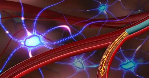
Straight (Top) and bended (Base) miniature endovascular (MEV) tests infused from microcatheters The film shows MEV tests intended to enter generally straight versus bended veins at branch focuses. Credit: Anqi Zhang, Stanford College.
In histology testing, the tests exhibited long-term soundness with negligible safe reactions. Tests couldn’t twist or infiltrate the vessel walls, causing no harm to the blood-mind obstruction, and didn’t altogether diminish the blood stream or cause neurologic shortfalls.
In vivo electrophysiology recording was effectively accomplished in the cortex and olfactory bulb of anesthetized rodents. The tests showed branch-particular implantation and activity, uncovering different terminating properties in neurological illness models. Single-unit movement recording was accomplished, showing single-cell goals across vessel walls.
The cerebrovasculature goes from huge, shallow cortical vessels to the microvasculature and slender beds inside the cortex. In the rodent mind, 5% of vessels have a measurement bigger than 100 m, which the review MEV tests could target.
Focusing on vessels of more modest breadth could be accomplished by decreasing the size and bowing firmness of the tests. At present, accessible endovascular tests for people and sheep have just had the option to focus on the biggest vessel above 2.4 mm in breadth.
The review creators infer that the “platform innovation could be stretched out to the discovery and treatment of numerous neurological sicknesses as an exploration instrument and could act as the establishment for clinical interpretation of negligibly obtrusive neuroelectronic interfaces.”
More information: Anqi Zhang et al, Ultraflexible endovascular probes for brain recording through micrometer-scale vasculature, Science (2023). DOI: 10.1126/science.adh3916


