Zeolites have one-of-a kind permeable nuclear designs and are valuable as impetuses, particle exchangers, and sub-atomic sifters. It is hard to straightforwardly notice the neighborhood nuclear designs of the material by means of electron microscopy because of the low electron illumination opposition. Thus, the key property-structure connections of the buildings stay indistinct.
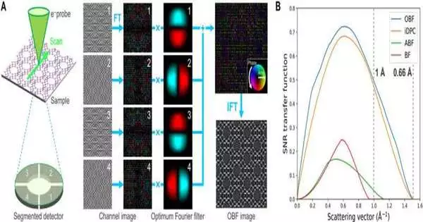
Ongoing improvements to a low-electron portion imaging strategy known as ideal splendid field checking transmission electron microscopy (OBF STEM) offer a technique to recreate pictures with a high sign-to-clamor proportion and high portion proficiency.
In this review, Kousuke Ooe and a group of researchers in design and nanoscience at the College of Tokyo and the Japan Fine Ceramics Community performed low-portion nuclear goal perceptions with the strategy to picture nuclear destinations and their systems between two kinds of zeolites. The researchers noticed the complex nuclear construction of the twin limits in a faujasite-type (FAU) zeolite to work with the portrayal of nearby nuclear designs across numerous electron-dense materials.
Breaking down zeolites in the materials lab
Zeolites are permeable materials that are consistently organized in nanosized pores appropriate for various applications during catalysis, gas partition, and particle trade. The material properties are firmly connected with the pore calculation, permitting ensuing cooperation with adsorbed visitor particles. Analysts have so far utilized diffractometric techniques to investigate the construction of zeolites.
For instance, materials researchers have shown that examining electron microscopy is a strong technique to dissect neighborhood designs to notice the nuclear plan of electron-safe materials at the sub-angstrom level. Zeolites are, be that as it may, more electron-bar delicate when contrasted with other natural materials, subsequently restricting electron microscopy-based perceptions because of electron illumination.
Ideal splendid field examining transmission electron microscopy (OBF/STEM)
In 1958, materials researcher J. W. Menter noticed zeolites utilizing a high-goal transmission electron magnifying lens to report a grid goal of 14 Angstrom. Pictures of the zeolite system significantly improved through cutting-edge imaging during the 1990s, in spite of the fact that it stayed too testing to notice the nuclear locales in the materials.
Late advances in filtering transmission electron microscopy (STEM) electron finders have prompted further development of imaging strategies like the ideal splendid field (OBF) STEM strategy to notice nuclear designs at the most noteworthy sign-to-clamor proportion to get nuclear goal pictures continuously.
In this work, Ooe and partners utilized continuous OBF imaging to determine the engineering of zeolites at the subangstrom goal. The results underlined the limit of cutting-edge electron microscopy to portray the nearby design of delicate materials.
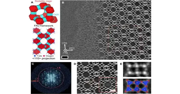
Nuclear goal OBF STEM perception of a FAU zeolite along the 110> zone pivot (A) Schematic of the FAU zeolite system structure and projected nuclear construction model along the 110 zone hub Red and blue polygons address the structure units (sodalite confines and D6Rs, individually). (B) OBF STEM picture of FAU zeolite seen at the edge of the example Brilliant spots demonstrate T and oxygen destinations. Scale bar, 1 nm The rectangle demonstrates the recurrent unit structure utilized for the averaging system displayed in (D). (C) Fourier change range of (B), wherein the spots are seen up to 0.869 Å goal in genuine space. (D) Rehash unit-cell-found the middle value of the OBF picture, which is gotten by trimming and averaging the various subimages got from the crude picture displayed in (B), offering a higher SNR. The inset is a recreated OBF picture determined with a similar perception condition as that in the examination. The area of the D6R structure, which is displayed in (E), is featured by a rectangle. (E) Amplified OBF picture of the rectangular locale shown by the red running line in (D) The nuclear construction models are drawn using representations for electronic and underlying investigation programming. Credit: Science Advances (2023). DOI: 10.1126/sciadv.adf6865
Direct imaging of nuclear designs in zeolites: Ongoing OBF imaging versus STEM imaging
The zeolite structure is comprised of two structural blocks: sodalite enclosures and twofold 6-membered rings. Utilizing continuous ideal brilliant field (OBF) imaging, the group identified the structure of the material and utilized an electron test current of 0.5 pico-angstrom to forestall any pillar-related harm in investigating the regular inorganic materials. They then, at that point, contrasted the OBF pictures with other checking transmission electron microscopy pictures acquired under comparable portion conditions.
The current STEM techniques showed a fundamental design of the material system; in any case, nuclear construction examination with this strategy was difficult because of the low current measurement. Conversely, the OBF pictures offered a more solid and interpretable picture that diverged from higher portion productivity.
Direct perception of the twin limit
The exploration group utilized the ideal splendid field technique to look at the nuclear design of a twin limit in the zeolite structure. The system was spread by cubic stacking a layered design unit as a “faujasite sheet.” The results of imaging with OBF showed a power range of the picture with data moving past 1 Angstrom. The low-portion light-component imaging with OBF STEM offered a superior choice to examine the construction of zeolites, including the neighborhood change of balance.
Ooe and partners used useful hypothesis estimations to inspect the soundness of the twin limit structure where the trial picture concurred with its mimicked partner.
The group applied the strategy to an alternate kind of zeolite test to show how the run-of-the mill silicon and aluminum proportions of these examples are urgent to the material properties and impact the adhesion of particles and atoms. At the point when they applied the technique to a sodium-based zeolite test for nuclear perceptions, the results showed the origination of additional cation locales with low inhabitance in the zeolitic system.
Standpoint
Along these lines, Kousuke Ooe and partners concocted a portion-effective filtering transmission electron microscopy imaging strategy known as “ideal splendid field examining transmission electron microscopy” (OBF-STEM) for low-portion nuclear goal imaging. The group showed how the technique straightforwardly uncovered the nuclear designs of all components in a faujasite-type zeolite material—a known pillar-touchy material with a subangstrom space goal.
The strategy can be utilized to recognize cross-sectional anomalies in the material system. They pictured the nuclear locales in the structure close by its caught cations to get results that were in quantitative concurrence with picture reenactments. The technique is pertinent across shafts of delicate materials past zeolites to portray the nearby nuclear construction and study the design property connections of delicate materials.
More information: Kousuke Ooe et al, Direct imaging of local atomic structures in zeolite using optimum bright-field scanning transmission electron microscopy, Science Advances (2023). DOI: 10.1126/sciadv.adf6865


