Beginning something new can be overwhelming if an individual does not understand what they will need or where to begin. Quantitative bioimaging, the method involved with getting quantitative information through pictures (some of the time a huge number of them), and computational methodologies, similar to AI, are generally new to the field of microscopy.
Quantitative bioimaging can be challenging for biologists without a background in computational science to incorporate into their experiments due to its novelty and complexity. Although there are resources to teach everything needed to perform quantitative bioimaging, the sheer quantity of educational materials can be overwhelming for those just starting out.
Beth Cimini, associate director for bioimage analysis in the Imaging Platform at the Broad Institute of MIT and Harvard, has created bioimagingguide.org, an online guide for producing high-quality microscopy images for quantitative data analysis, in collaboration with BioImaging North America, an organization that encourages collaboration in the bioimaging community.
With the aide and a buddy paper distributed in PLOS Science, Cimini, postdoctoral colleagues Rebecca Senft and Barbara Diaz-Rohrer, and their partners plan to make an asset that makes the devices, strategies, and standards of quantitative bioimaging more open to scientists who don’t have a computational foundation.
We sat down with Cimini to talk about what bioimagingguide.org is and the way in which it can assist researchers who need to bring bioimaging into their exploration.
What is the underlying concept of bioimagingguide.org?
Good quantitative microscopy requires many steps, and there is a lot to learn about each one. A ton of spots run perfectly-escalated microscopy courses, yet those courses are held for around 20 individuals every year. What’s more, perhaps you’re sufficiently fortunate to work at a spot with an incredible microscopy community where they run a course like this consistently. Many individuals aren’t, in any case.
We felt that there was a “big picture” overview of microscopy that wasn’t a full textbook but rather a medium-sized guide that said, “Here are all the things you have to learn, and here are links to great resources that we or other people have written to learn more.” This was what we felt was missing.
So our objective was to assemble something that gives an exhaustive diagram of the points one has to be aware of to get everything rolling in bioimaging and which connections individuals have to data that they can reference at their own speed.
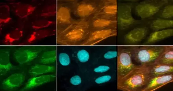
A collection portraying a solitary cell test imaged in various channels utilizing the Cell Painting convention. Credit: Beth Cimini, Imaging Platform
The aid initially began as only a paper, yet we soon understood that there was an excess of data. Likewise, we believed we should add new things as we track them down and keep up with the latest. A paper is a static moment and shows what data was accessible when it was delivered. We wanted to be able to bring in a great new piece if someone wrote it tomorrow.
What sorts of bioimaging tests did you have as a primary concern while composing the aide?
Bioimaging examinations can go from a solitary picture where somebody needs to quantify one thing to terabytes of pictures where they need to gauge thousands. The person who needs to stain a sample, take a few dozen fluorescent images, and learn two or three things from those images is the target audience for a lot of the information in the guide.
How is this guide not the same as different assets?
This guide is novel in that it shows the significant subjects of bioimaging to fledglings in an open, online configuration that unites and coordinates a lot of content that generally existed somewhere else. We purposefully organized it so that individuals could ingest data at an expansive level, yet assuming they need subtleties, they can undoubtedly track down more data. Not all things are introduced on the screen on the double, which I trust can make it less overpowering to begin with. Rebecca and Barbara worked really hard, driving about 15 creators on the paper and assembling a few extraordinary assets.
Why did you all decide to write the guide?
In the Imaging Stage, we make open-source devices and work processes for scientists at Wide and then some. Overall, we team up with 20 to 30 gatherings a year on tasks and converse with many specialists. In conversing with these people, we frequently hear similar disappointments about rehashing portions of a task in view of blunders that might have been avoided if they had known what to do.
We had already planned to develop a method to assist individuals in generating accurate data suitable for accurate quantification, and during these discussions, we realized that an introduction to effective microscopy was also required. As a result, we contacted the Image Informatics Working Group of BioImaging North America, which focuses on image analysis, and the Training and Education Working Group, which focuses on microscopy education, to seek their guidance in the development of a project similar to this one.
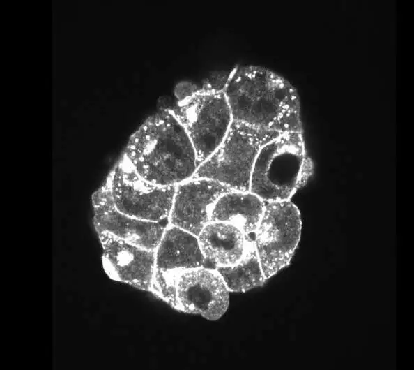
A heap of mouse trophoblast immature microorganisms was imaged utilizing fluorescent microscopy. Credit: Rivron et al. 2018, available from the Broad Bioimage Benchmark Collection [Ljosa et al., Nature Methods, 2012]
What is your number one component on the site?
There are a great deal of elements to cherish, but the fact that practically all the data in the aid is accessible first rings a bell. In addition to a glossary of terms and definitions, there are numerous links to papers and other websites.
I’m likewise truly amped up for an element that we’re dealing with yet that isn’t yet live: website translation done automatically. Few out of every odd researcher is conversant in English, and keeping in mind that somebody can constantly utilize Google Decipher or something to that effect, such interpretations are noticeably flawed all the time. In addition to translating all of the content into French, Spanish, and Portuguese with the Training and Education Working Group, we are collaborating with others to translate the website into additional languages.
How would you expect or trust that individuals will utilize this aid?
All the time, you’ll find quantitative bioimaging doesn’t occur straight away, but rather all around. An individual could arrive at a point in their undertaking and understand that something they did beforehand has made it so they can’t continue. They should then return to the seat and attempt an alternate immune response or color, or even an alternate sort of magnifying instrument.
My hope is that the data we’ve gathered will help people figure out where they are on that circle and get to the answers they want more quickly.
More information: Rebecca A. Senft et al, A biologist’s guide to planning and performing quantitative bioimaging experiments, PLOS Biology (2023). DOI: 10.1371/journal.pbio.3002167


