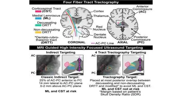
UT Southwestern doctors have fostered an improved focus on strategy, four-lot tractography, to customize X-ray-directed, extreme focus centered ultrasound (HIFU) utilized at UTSW to treat drug-resistant tremor in the fundamental, endlessly recurring tremor of Parkinson’s illness. The aftereffects of the clinical cases using this methodology, distributed in Mind Correspondences, propose that it empowers more exact focusing of the cerebrum, diminishes treatment times, lessens secondary effects, and further develops the treatment reaction.
“By and large, the quake’s target remained invisible on standard high-goal attractive reverberation images, so milestone-based techniques were used,” said Bhavya R. Shah, M.D., assistant professor of radiology and neurological medical procedure, agent in the Peter O’Donnell Jr. Mind Foundation, and relating creator.”By utilizing tractography and other high-level attractive reverberation strategies, we are attempting to recognize the exact area of the objective in individual patients and work toward quiet results.”
X-ray-directed HIFU removal of the ventral middle core of the thalamus has been endorsed by the Food and Drug Administration for treatment of fundamentally irreversible Parkinson’s disease. The adequacy and security of the system are subject to exact focusing; in any case, X-rays can’t straightforwardly depict the objective. Hence, aberrant techniques utilizing milestones are used to recognize the right locale.
“Historically, the target for tremor could not be visible on typical high-resolution magnetic resonance images, hence landmark-based approaches were used. We are combining tractography and other advanced magnetic resonance approaches to pinpoint the precise position of the target in individual patients and improve patient outcomes.”
Bhavya R. Shah, M.D., assistant professor of radiology and neurological surgery,
The use of circuitous focusing for HIFU removal can cause significant unfriendly secondary effects, including shortcoming, death, and paresthesia in nearly 33% of patients one month after treatment, which can be alleviated by utilizing methods to more directly focus on the area of the mind and recognize areas to avoid.
To that end, the UT Southwestern researchers developed an improved, standardized four-lot tractography for HIFU using an FDA-supported careful planning program that will take into account simple execution in the centers.

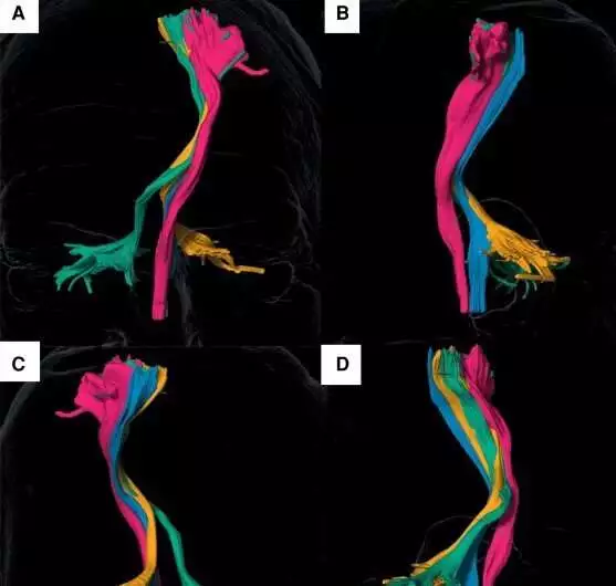
Four-lot tractography recognizes white-matter plots in the mind, displayed in 3D renderings, for therapy with X-ray-directed, extreme focus-centered ultrasound (HIFU).
The four-lot tractography strategy works off past work by Dr. Shah, distributed in Mind in 2020, in which the analysts showed the possibility of cutting-edge X-ray methods to recognize quake-related white matter lots.
The most recent review looked at data from 18 patients who had X-ray-directed HIFU and had at least 90 days of follow-up after treatment.
Utilization of the four-lot tractography for removal suggests that this strategy yields diminished unfriendly impacts. After treatment, none of the patients experienced shortcoming, deadness, or paresthesia; three patients experienced transient (three-week) unevenness that resolved after attractive reverberation-directed HIFU.
However, this review survey was restricted by the small number of patients treated in one place with a short development, but the outcomes proposed a guarantee for four-lot tractography to create improved results for patients who needed this method. A multicenter preliminary is arranged with other U.S. locales, including the College of California, San Francisco, the Mayo Center in Rochester, Minnesota, the College of Colorado, and a global site in Spain.
A few different locales have organized to come to UTSW to work with Dr. Shah and get familiar with his methodology. According to Dr. Shah, this work is a step in the right direction because it uses advanced imaging to provide customized answers for these patients.
More information: Fabricio S Feltrin et al, Focused ultrasound using a novel targeting method four-tract tractography for magnetic resonance–guided high-intensity focused ultrasound targeting, Brain Communications (2022). DOI: 10.1093/braincomms/fcac273
Journal information: Brain Communications


