The mind is seemingly quite possibly of the most complicated structure in the known universe.
Proceeded with progresses in how we might interpret the cerebrum and our capacity to really treat a large group of neurological illnesses depend on testing the mind’s brain miniature hardware with steadily expanding subtlety.
One class of strategies for concentrating on brain circuits is called voltage imaging. These strategies permit us to see the voltage created by our cerebrum’s terminating neurons — letting us know how organizations of neurons create, capability and change after some time.
Today, voltage imaging of refined neurons is performed utilizing thick varieties of anodes onto which cells are developed (or refined), or by applying light-discharging colors that answer optically to changes in voltage on the outer layer of the phone.
In any case, the degree of detail we can see utilizing these procedures is confined.
The littlest terminals can’t dependably recognize individual neurons, exactly 20 millionths of a meter across, to not express anything of the thick organization of nanoscale associations that structures among them, and no huge mechanical advances have been made around here for more than twenty years.
Moreover, every terminal requires its own wired association and speaker, putting critical constraints on the quantity of cathodes that can be estimated at the same time.
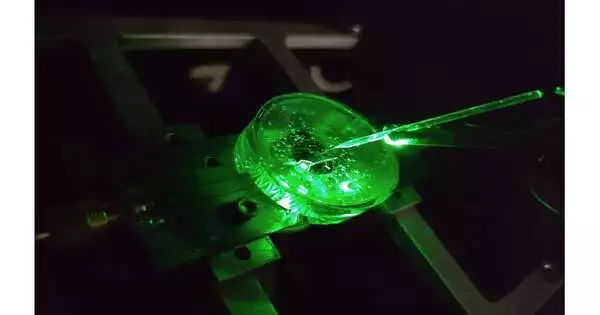
A minuscule terminal, eight millionths of a meter across, is utilized to locally infuse a haze of electrical surge into a fluid set on the precious stone chip. The fluorescence from the precious stone mirrors the dissemination of this charge through the fluid continuously. Credit: Creator gave
Colors can defeat these impediments by imaging the voltage remotely as light — this implies the mind boggling gadgets can be arranged away from the cells inside a camera.
The outcome is high goal over huge regions, ready to recognize every individual neuron in an enormous organization. In any case, there are constraints here as well, the voltage reactions of best in class colors are slow and unsteady.
Our new exploration distributed in Nature Photonics, investigates another sort of a high velocity, high goal and versatile voltage imaging stage made fully intent on beating these restrictions — a precious stone voltage imaging magnifying lens.
Created by a group of physicists from the College of Melbourne and RMIT College, the gadget utilizes a precious stone based sensor that changes over voltage signals at its surface straightforwardly into optical signs — this implies we can see electrical movement as it works out.
The change utilizes the properties of an iota scale imperfection in the jewel’s gem structure known as the nitrogen-opening (NV).
NV imperfections can be designed by barraging the jewel with a nitrogen particle pillar utilizing an exceptional sort of atom smasher. The manufacture of the sensor starts with utilizing this cycle to make a high-thickness, super flimsy layer of NV surrenders near the precious stone’s surface.
You can imagine every NV imperfection as a pail that holds up to two electrons. At the point when this pail is unfilled, the NV deformity is dull. With one electron, the NV imperfection produces orange light when enlightened by a laser — this property is known as fluorescence. With two electrons, the shade of the fluorescence becomes red.
As the voltage in a conductive arrangement is consistently changed, the brilliance of light discharged by the precious stone chip follows with a close to prompt reaction. Here, the jewel surface has been designed into a variety of nanopillars to expand the recognized light sign. Credit: Creator gave
A formerly found property of NV deserts is that the quantity of electrons they hold — and the subsequent fluorescence — can be controlled with a voltage. Not at all like colors, the voltage reaction of a NV deformity is extremely quick and stable.
Our examination plans to conquer the test of making this impact adequately delicate to picture neuronal movement.
On the jewel’s surface, the precious stone construction closes with a layer one particle thick, comprised of hydrogen and oxygen iotas. The NV deserts nearest to the surface are the most delicate to changes in voltage outside the jewel, however they are additionally exceptionally delicate to the nuclear cosmetics of the surface layer.
An excessive amount of hydrogen and the NVs are dim to such an extent that the optical signs we are searching for shouldn’t be visible. Too little hydrogen and the NVs are splendid to such an extent that the little signals we many ares totally cleaned out.
Thus, there’s a “Goldilocks’ zone” for voltage imaging, where the surface has a perfect proportion of hydrogen.
To arrive at this zone, our group fostered an electrochemical strategy for eliminating hydrogen in a controlled manner. By doing this, we’ve figured out how to accomplish voltage responsive qualities two significant degrees better than whatever has been recently detailed.
We tried our sensor in pungent water utilizing a minute wire 10-times more slender than a human hair. By applying a momentum, the wire can create a little haze of charge in the water over the precious stone. The development and ensuing dissemination of this charge cloud creates little voltages at the jewel surface.
By catching these voltages through a rapid recording of the NV fluorescence, we can decide the speed, responsiveness and goal of our jewel imaging chip.
We had the option to additional lift responsiveness by designing the jewel’s surface into ‘nanopillars’ — funnel shaped structures with the NV habitats implanted in their tips. These points of support channel the light discharged by the NVs towards the camera, decisively expanding how much sign we can gather.
With the advancement of the precious stone voltage imaging magnifying lens for identifying neuronal action, the subsequent stage is the recording of action from refined neurons in vitro — these are probes cells developed external their typical natural setting, also called test-tube or petri-dish tests.
What separates this innovation from existing best in class in vitro procedures is the blend of high spatial goal (on the request for a millionth of a meter or less), huge spatial scale (a couple of millimeters toward every path — which for an organization of neurons in warm blooded creatures is very tremendous), and complete steadiness over the long run.
No other existing framework can at the same time offer these three characteristics, and this blend will permit our made-in-Melbourne innovation to worldwide make an important commitment to crafted by neuroscientists and neuropharmacologists.
Our framework will help these scientists in seeking after both crucial information and the up and coming age of medicines for neurological and neurodegenerative sicknesses.
More information: D. J. McCloskey et al, A diamond voltage imaging microscope, Nature Photonics (2022). DOI: 10.1038/s41566-022-01064-1
Journal information: Nature Photonics


