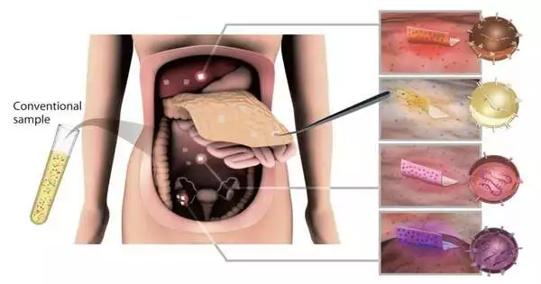
An exploration group in Japan, led by Nagoya College’s Akira Yokoi, has fostered a creative method utilizing cellulose nanofiber (CNF) sheets made from wood cellulose to catch extracellular vesicles (EVs) from liquid examples and even organs during medical procedures.
EVs are little designs from harmful cells that assume an urgent role in cell-to-cell correspondence. Extricating and investigating EVs utilizing this new innovation can possibly upset early malignant growth determination and pave the way for customized medication. The scientists have distributed their discoveries in Nature correspondences.
Disease is famous for its unfortunate anticipation, and much of the time it goes undetected until its high-level stages, leaving patients with restricted treatment choices. Identifying the malignant growth early, utilizing EVs, and investigating them gives indispensable data on infection status and its movement. This ought to help doctors observe and change customized disease treatment plans. In any case, specialists have been restricted in past endeavors to utilize EVs because of the absence of a viable segregation procedure.
“We created the one-of-a-kind cellulose nanofiber by combining papermaking and solvent displacement technology. The cellulose nanofibers we use are a sustainable biomass material derived primarily from wood cell walls. These sheets offer appealing characteristics such as being lightweight, strong, and, most significantly, easily biodegradable.”
Nagoya University’s Akira Yokoi,
To catch EVs, Yokoi and his partners utilized CNF sheets produced using wood cellulose to extricate them from liquid examples from ovarian disease mouse models. As the material has a permeable nanostructure, the sheet ingests the liquid containing the EVs into its pores and closes them after drying. They found that the sheets caught and saved EVs from just ten microliters of body liquid. Interestingly, the ongoing standard techniques, like ultra-centrifugation, are extra tedious and require a lot more examples.
“We have fostered the interesting cellulose nanofiber by applying paper production and dissolvable dislodging innovation,” Yokoi said. “The cellulose nanofibers we use are a manageable biomass material that comes generally from wood cell walls. These sheets have alluring properties, for example, being lightweight, high-strength, and, above all, effectively biodegradable.”
Utilizing the strategy, the scientists effectively extricated and dissected EVs and the microRNAs (miRNAs) held inside them from the mouse ovarian disease models. As miRNAs vary among sound and debilitated patients, they address an optimal indicative marker for disease. The group likewise recognized unmistakable arrangements of miRNAs in EVs gathered from growth surfaces, some of which diminished after cancer expulsion.
Following the presence or nonattendance of these miRNAs could be a simple method for investigating the adequacy of treatment and then tailoring treatment as indicated by growth heterogeneity. Heterogeneity is a typical issue where, even in solitary growth, disease cells have various qualities and properties.
The construction of the sheets is like that of clinical cloth, so they can undoubtedly be joined and eliminated in any event when put on organs during a medical procedure. To test this, the gathering utilized as of late has taken out human organs. Their fruitful test uncovered a thrilling revelation, as the EVs on the cancer surface showed novel miRNA profiles that contrasted with the growth tissue.
“Organ surfaces were a formerly unanalyzed EV subpopulation, which can now be exposed to natural appraisals,” Yokoi said. “CNF paper empowers the getting of EVs from numerous destinations in the body. Then, by checking the sub-atomic profiles of these EVs, we can screen sickness movement and design the choice of the best medication, adding to customized medication.”
Dr. Takahiro Ochiya, Board Individual from the Global Society for Extracellular Vesicles and Leader of the Japanese Society for Extracellular Vesicles, is energetic about the capability of the sheets, saying, “Exosome investigation utilizing CNF sheets is an incredibly original strategy and is supposed to have different applications, including clinical purposes. We anticipate that this should be a serious step forward that will bring the information on exosomes as clinical exploration straightforwardly to patients.”
This exploration has expansive ramifications, opening up the examination of EVs during medical procedures, a neglected region as of recently. Looking forward, the exploration group is focused on propelling the clinical utilization of EV sheets for different illnesses, working on demonstrative exactness, and introducing the time of customized medication.
More information: Spatial exosome analysis using cellulose nanofiber sheets reveals the location heterogeneity of extracellular vesicles, Nature Communications (2023).


